Copper »
PDB 3mnd-3qjo »
3phm »
Copper in PDB 3phm: Reduced (Cu+) Peptidylglycine Alpha-Hydroxylating Monooxygenase (Phm)
Enzymatic activity of Reduced (Cu+) Peptidylglycine Alpha-Hydroxylating Monooxygenase (Phm)
All present enzymatic activity of Reduced (Cu+) Peptidylglycine Alpha-Hydroxylating Monooxygenase (Phm):
1.14.17.3;
1.14.17.3;
Protein crystallography data
The structure of Reduced (Cu+) Peptidylglycine Alpha-Hydroxylating Monooxygenase (Phm), PDB code: 3phm
was solved by
S.T.Prigge,
L.M.Amzel,
with X-Ray Crystallography technique. A brief refinement statistics is given in the table below:
| Resolution Low / High (Å) | 10.00 / 2.10 |
| Space group | P 21 21 21 |
| Cell size a, b, c (Å), α, β, γ (°) | 69.519, 68.770, 82.417, 90.00, 90.00, 90.00 |
| R / Rfree (%) | 20 / 25.7 |
Other elements in 3phm:
The structure of Reduced (Cu+) Peptidylglycine Alpha-Hydroxylating Monooxygenase (Phm) also contains other interesting chemical elements:
| Nickel | (Ni) | 1 atom |
Copper Binding Sites:
The binding sites of Copper atom in the Reduced (Cu+) Peptidylglycine Alpha-Hydroxylating Monooxygenase (Phm)
(pdb code 3phm). This binding sites where shown within
5.0 Angstroms radius around Copper atom.
In total 2 binding sites of Copper where determined in the Reduced (Cu+) Peptidylglycine Alpha-Hydroxylating Monooxygenase (Phm), PDB code: 3phm:
Jump to Copper binding site number: 1; 2;
In total 2 binding sites of Copper where determined in the Reduced (Cu+) Peptidylglycine Alpha-Hydroxylating Monooxygenase (Phm), PDB code: 3phm:
Jump to Copper binding site number: 1; 2;
Copper binding site 1 out of 2 in 3phm
Go back to
Copper binding site 1 out
of 2 in the Reduced (Cu+) Peptidylglycine Alpha-Hydroxylating Monooxygenase (Phm)
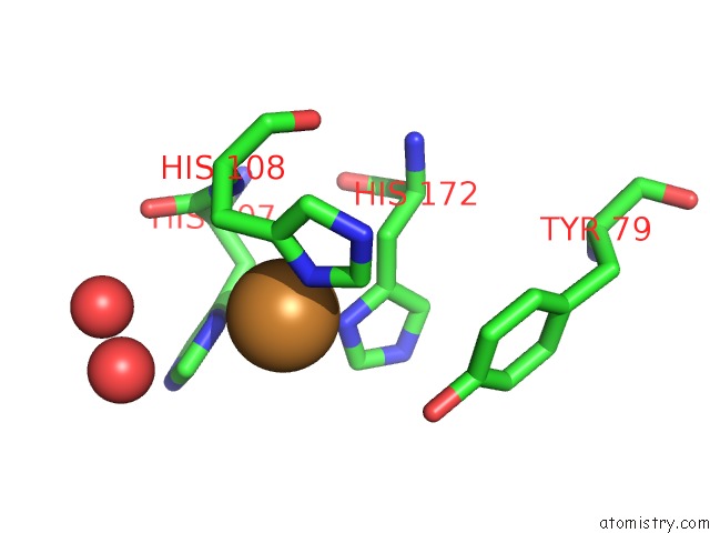
Mono view
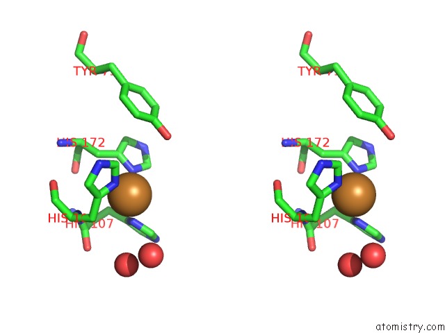
Stereo pair view
Mono view
Stereo pair view
A full contact list of Copper with other atoms in the Cu binding
site number 1 of Reduced (Cu+) Peptidylglycine Alpha-Hydroxylating Monooxygenase (Phm) within 5.0Å range:
|
Copper binding site 2 out of 2 in 3phm
Go back to
Copper binding site 2 out
of 2 in the Reduced (Cu+) Peptidylglycine Alpha-Hydroxylating Monooxygenase (Phm)
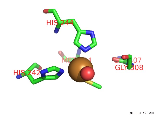
Mono view
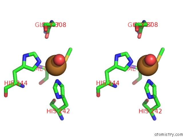
Stereo pair view
Mono view
Stereo pair view
A full contact list of Copper with other atoms in the Cu binding
site number 2 of Reduced (Cu+) Peptidylglycine Alpha-Hydroxylating Monooxygenase (Phm) within 5.0Å range:
|
Reference:
S.T.Prigge,
A.S.Kolhekar,
B.A.Eipper,
R.E.Mains,
L.M.Amzel.
Substrate-Mediated Electron Transfer in Peptidylglycine Alpha-Hydroxylating Monooxygenase. Nat.Struct.Biol. V. 6 976 1999.
ISSN: ISSN 1072-8368
PubMed: 10504734
DOI: 10.1038/13351
Page generated: Mon Jul 14 02:35:50 2025
ISSN: ISSN 1072-8368
PubMed: 10504734
DOI: 10.1038/13351
Last articles
Mn in 4XISMn in 4XBW
Mn in 4X9Q
Mn in 4WV8
Mn in 4X8D
Mn in 4WZQ
Mn in 4WZM
Mn in 4WYL
Mn in 4WUO
Mn in 4WTM