Copper »
PDB 3mnd-3qjo »
3n7e »
Copper in PDB 3n7e: Crystal Structure of Copk Bound to Cu(II)
Protein crystallography data
The structure of Crystal Structure of Copk Bound to Cu(II), PDB code: 3n7e
was solved by
M.-R.Ash,
M.J.Maher,
with X-Ray Crystallography technique. A brief refinement statistics is given in the table below:
| Resolution Low / High (Å) | 46.42 / 2.30 |
| Space group | C 1 2 1 |
| Cell size a, b, c (Å), α, β, γ (°) | 49.835, 51.593, 52.054, 90.00, 116.91, 90.00 |
| R / Rfree (%) | 19.9 / 26.7 |
Copper Binding Sites:
The binding sites of Copper atom in the Crystal Structure of Copk Bound to Cu(II)
(pdb code 3n7e). This binding sites where shown within
5.0 Angstroms radius around Copper atom.
In total 2 binding sites of Copper where determined in the Crystal Structure of Copk Bound to Cu(II), PDB code: 3n7e:
Jump to Copper binding site number: 1; 2;
In total 2 binding sites of Copper where determined in the Crystal Structure of Copk Bound to Cu(II), PDB code: 3n7e:
Jump to Copper binding site number: 1; 2;
Copper binding site 1 out of 2 in 3n7e
Go back to
Copper binding site 1 out
of 2 in the Crystal Structure of Copk Bound to Cu(II)
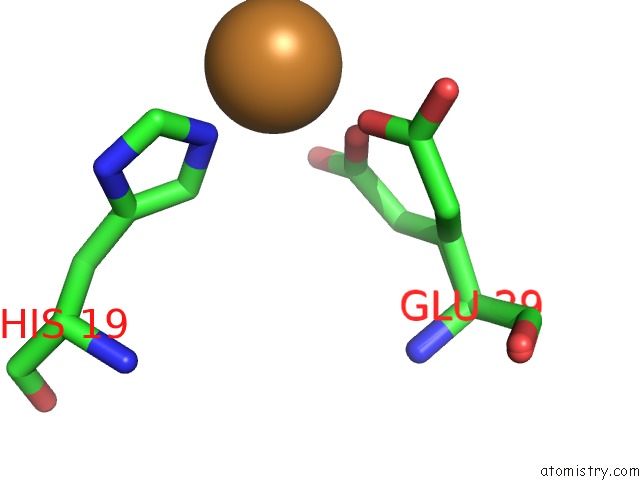
Mono view
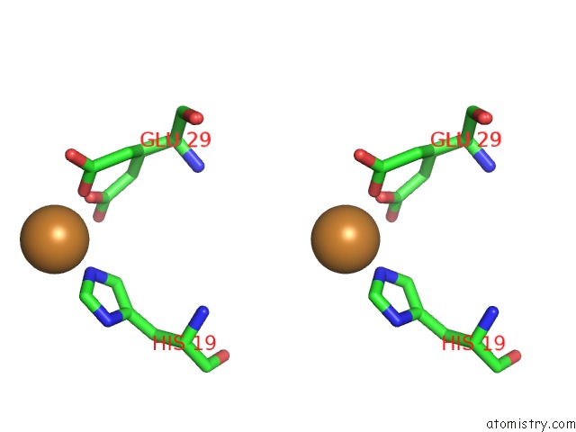
Stereo pair view
Mono view
Stereo pair view
A full contact list of Copper with other atoms in the Cu binding
site number 1 of Crystal Structure of Copk Bound to Cu(II) within 5.0Å range:
|
Copper binding site 2 out of 2 in 3n7e
Go back to
Copper binding site 2 out
of 2 in the Crystal Structure of Copk Bound to Cu(II)
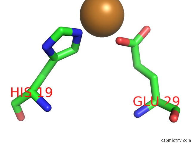
Mono view
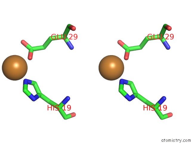
Stereo pair view
Mono view
Stereo pair view
A full contact list of Copper with other atoms in the Cu binding
site number 2 of Crystal Structure of Copk Bound to Cu(II) within 5.0Å range:
|
Reference:
M.-R.Ash,
M.J.Maher.
Two New Crystal Forms of Copper Resistance Protein Copk To Be Published.
Page generated: Mon Jul 14 02:26:52 2025
Last articles
I in 4B9HI in 4AS2
I in 4AS5
I in 4AX2
I in 4ARR
I in 4AQ3
I in 4ARK
I in 4AP2
I in 4AIO
I in 4ANB