Copper »
PDB 3awt-3erx »
3dso »
Copper in PDB 3dso: Crystal Structure of Cu(I) Bound Copper Resistance Protein Copk
Protein crystallography data
The structure of Crystal Structure of Cu(I) Bound Copper Resistance Protein Copk, PDB code: 3dso
was solved by
M.-R.Ash,
M.J.Maher,
with X-Ray Crystallography technique. A brief refinement statistics is given in the table below:
| Resolution Low / High (Å) | 15.80 / 1.55 |
| Space group | P 21 21 2 |
| Cell size a, b, c (Å), α, β, γ (°) | 32.193, 84.672, 23.662, 90.00, 90.00, 90.00 |
| R / Rfree (%) | 20.3 / 21.2 |
Copper Binding Sites:
The binding sites of Copper atom in the Crystal Structure of Cu(I) Bound Copper Resistance Protein Copk
(pdb code 3dso). This binding sites where shown within
5.0 Angstroms radius around Copper atom.
In total only one binding site of Copper was determined in the Crystal Structure of Cu(I) Bound Copper Resistance Protein Copk, PDB code: 3dso:
In total only one binding site of Copper was determined in the Crystal Structure of Cu(I) Bound Copper Resistance Protein Copk, PDB code: 3dso:
Copper binding site 1 out of 1 in 3dso
Go back to
Copper binding site 1 out
of 1 in the Crystal Structure of Cu(I) Bound Copper Resistance Protein Copk
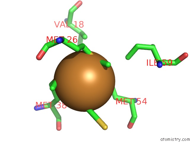
Mono view
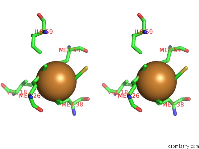
Stereo pair view
Mono view
Stereo pair view
A full contact list of Copper with other atoms in the Cu binding
site number 1 of Crystal Structure of Cu(I) Bound Copper Resistance Protein Copk within 5.0Å range:
|
Reference:
L.X.Chong,
M.-R.Ash,
M.J.Maher,
M.G.Hinds,
Z.Xiao,
A.G.Wedd.
Unprecedented Binding Cooperativity Between Cu(I) and Cu(II) in the Copper Resistance Protein Copk From Cupriavidus Metallidurans CH34: Implications From Structural Studies By uc(Nmr) Spectroscopy and X-Ray Crystallography J.Am.Chem.Soc. V. 131 3549 2009.
ISSN: ISSN 0002-7863
PubMed: 19236095
DOI: 10.1021/JA807354Z
Page generated: Mon Jul 14 02:02:47 2025
ISSN: ISSN 0002-7863
PubMed: 19236095
DOI: 10.1021/JA807354Z
Last articles
K in 8CGRK in 8CGJ
K in 8CGI
K in 8CG1
K in 8CFY
K in 8CG2
K in 8CFX
K in 8CG0
K in 8CFW
K in 8CFV