Copper »
PDB 3awt-3erx »
3azu »
Copper in PDB 3azu: X-Ray Crystal Structure of the Two Site-Specific Mutants HIS35GLN and HIS35LEU of Azurin From Pseudomonas Aeruginosa
Protein crystallography data
The structure of X-Ray Crystal Structure of the Two Site-Specific Mutants HIS35GLN and HIS35LEU of Azurin From Pseudomonas Aeruginosa, PDB code: 3azu
was solved by
A.Messerschmidt,
H.Nar,
R.Huber,
with X-Ray Crystallography technique. A brief refinement statistics is given in the table below:
| Resolution Low / High (Å) | N/A / 2.10 |
| Space group | P 21 21 21 |
| Cell size a, b, c (Å), α, β, γ (°) | 109.740, 99.150, 47.820, 90.00, 90.00, 90.00 |
| R / Rfree (%) | 16.3 / n/a |
Copper Binding Sites:
The binding sites of Copper atom in the X-Ray Crystal Structure of the Two Site-Specific Mutants HIS35GLN and HIS35LEU of Azurin From Pseudomonas Aeruginosa
(pdb code 3azu). This binding sites where shown within
5.0 Angstroms radius around Copper atom.
In total 4 binding sites of Copper where determined in the X-Ray Crystal Structure of the Two Site-Specific Mutants HIS35GLN and HIS35LEU of Azurin From Pseudomonas Aeruginosa, PDB code: 3azu:
Jump to Copper binding site number: 1; 2; 3; 4;
In total 4 binding sites of Copper where determined in the X-Ray Crystal Structure of the Two Site-Specific Mutants HIS35GLN and HIS35LEU of Azurin From Pseudomonas Aeruginosa, PDB code: 3azu:
Jump to Copper binding site number: 1; 2; 3; 4;
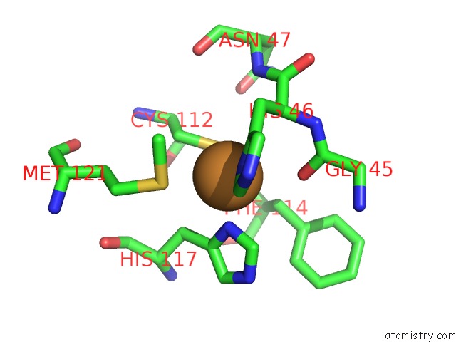
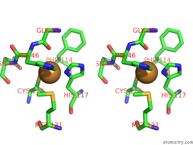
Copper binding site 1 out of 4 in 3azu
Go back to
Copper binding site 1 out
of 4 in the X-Ray Crystal Structure of the Two Site-Specific Mutants HIS35GLN and HIS35LEU of Azurin From Pseudomonas Aeruginosa
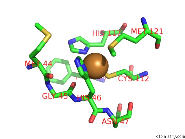
Mono view
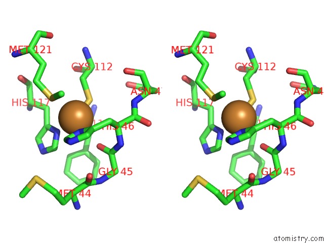
Stereo pair view
Mono view
Stereo pair view
A full contact list of Copper with other atoms in the Cu binding
site number 1 of X-Ray Crystal Structure of the Two Site-Specific Mutants HIS35GLN and HIS35LEU of Azurin From Pseudomonas Aeruginosa within 5.0Å range:
|
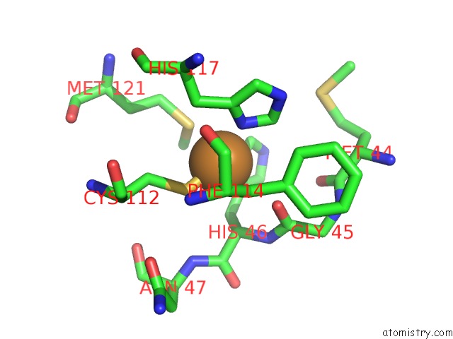
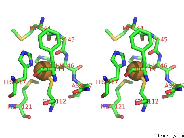
Copper binding site 2 out of 4 in 3azu
Go back to
Copper binding site 2 out
of 4 in the X-Ray Crystal Structure of the Two Site-Specific Mutants HIS35GLN and HIS35LEU of Azurin From Pseudomonas Aeruginosa
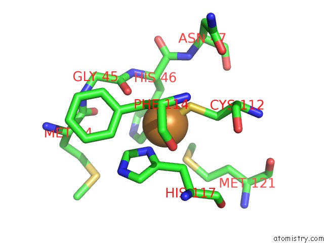
Mono view
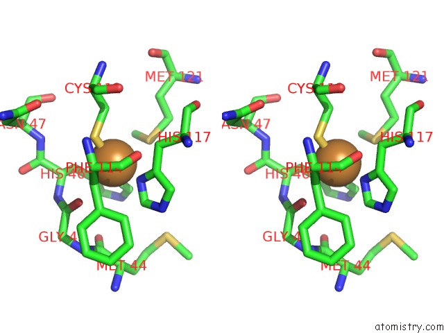
Stereo pair view
Mono view
Stereo pair view
A full contact list of Copper with other atoms in the Cu binding
site number 2 of X-Ray Crystal Structure of the Two Site-Specific Mutants HIS35GLN and HIS35LEU of Azurin From Pseudomonas Aeruginosa within 5.0Å range:
|
Copper binding site 3 out of 4 in 3azu
Go back to
Copper binding site 3 out
of 4 in the X-Ray Crystal Structure of the Two Site-Specific Mutants HIS35GLN and HIS35LEU of Azurin From Pseudomonas Aeruginosa
Mono view
Stereo pair view
Mono view
Stereo pair view
A full contact list of Copper with other atoms in the Cu binding
site number 3 of X-Ray Crystal Structure of the Two Site-Specific Mutants HIS35GLN and HIS35LEU of Azurin From Pseudomonas Aeruginosa within 5.0Å range:
|
Copper binding site 4 out of 4 in 3azu
Go back to
Copper binding site 4 out
of 4 in the X-Ray Crystal Structure of the Two Site-Specific Mutants HIS35GLN and HIS35LEU of Azurin From Pseudomonas Aeruginosa
Mono view
Stereo pair view
Mono view
Stereo pair view
A full contact list of Copper with other atoms in the Cu binding
site number 4 of X-Ray Crystal Structure of the Two Site-Specific Mutants HIS35GLN and HIS35LEU of Azurin From Pseudomonas Aeruginosa within 5.0Å range:
|
Reference:
H.Nar,
A.Messerschmidt,
R.Huber,
M.Van De Kamp,
G.W.Canters.
X-Ray Crystal Structure of the Two Site-Specific Mutants HIS35GLN and HIS35LEU of Azurin From Pseudomonas Aeruginosa. J.Mol.Biol. V. 218 427 1991.
ISSN: ISSN 0022-2836
PubMed: 1901363
DOI: 10.1016/0022-2836(91)90723-J
Page generated: Mon Jul 14 01:57:54 2025
ISSN: ISSN 0022-2836
PubMed: 1901363
DOI: 10.1016/0022-2836(91)90723-J
Last articles
K in 6QD4K in 6QM2
K in 6QD3
K in 6QD2
K in 6QD1
K in 6Q6R
K in 6QD0
K in 6QCZ
K in 6QCY
K in 6Q8P