Copper »
PDB 8uem-9cst »
9bjr »
Copper in PDB 9bjr: X-Ray Crystal Structure of Y168F Variant Thermothelomyces Thermophilus Polysaccharide Monooxygenase 9E
Protein crystallography data
The structure of X-Ray Crystal Structure of Y168F Variant Thermothelomyces Thermophilus Polysaccharide Monooxygenase 9E, PDB code: 9bjr
was solved by
W.C.Thomas,
R.I.Sayler,
M.A.Marletta,
with X-Ray Crystallography technique. A brief refinement statistics is given in the table below:
| Resolution Low / High (Å) | 58.62 / 2.20 |
| Space group | C 2 2 21 |
| Cell size a, b, c (Å), α, β, γ (°) | 97.989, 119.97, 92.29, 90, 90, 90 |
| R / Rfree (%) | 17.5 / 21.3 |
Copper Binding Sites:
The binding sites of Copper atom in the X-Ray Crystal Structure of Y168F Variant Thermothelomyces Thermophilus Polysaccharide Monooxygenase 9E
(pdb code 9bjr). This binding sites where shown within
5.0 Angstroms radius around Copper atom.
In total 2 binding sites of Copper where determined in the X-Ray Crystal Structure of Y168F Variant Thermothelomyces Thermophilus Polysaccharide Monooxygenase 9E, PDB code: 9bjr:
Jump to Copper binding site number: 1; 2;
In total 2 binding sites of Copper where determined in the X-Ray Crystal Structure of Y168F Variant Thermothelomyces Thermophilus Polysaccharide Monooxygenase 9E, PDB code: 9bjr:
Jump to Copper binding site number: 1; 2;
Copper binding site 1 out of 2 in 9bjr
Go back to
Copper binding site 1 out
of 2 in the X-Ray Crystal Structure of Y168F Variant Thermothelomyces Thermophilus Polysaccharide Monooxygenase 9E

Mono view
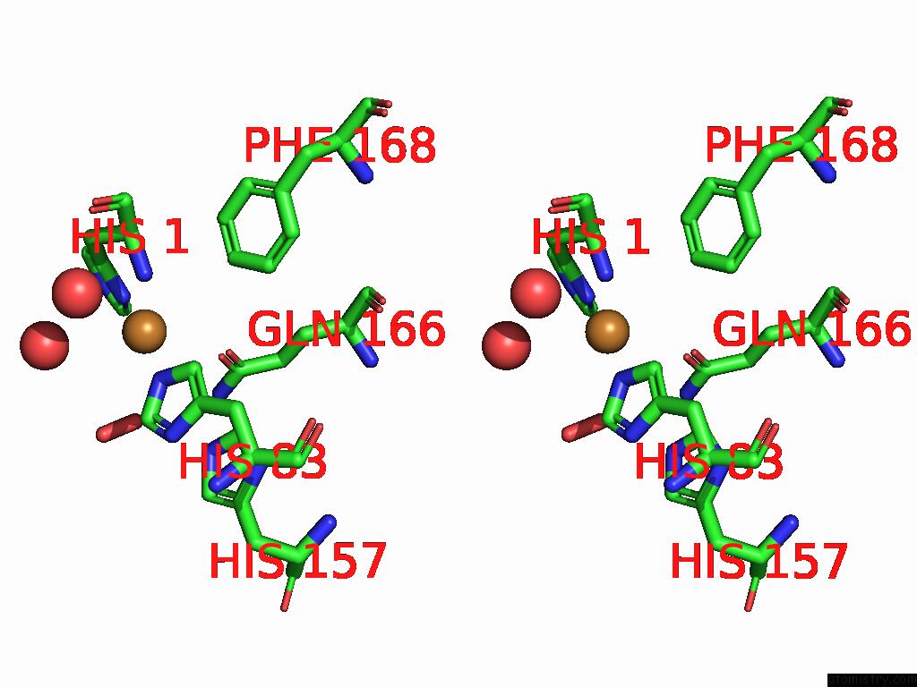
Stereo pair view
Mono view
Stereo pair view
A full contact list of Copper with other atoms in the Cu binding
site number 1 of X-Ray Crystal Structure of Y168F Variant Thermothelomyces Thermophilus Polysaccharide Monooxygenase 9E within 5.0Å range:
|
Copper binding site 2 out of 2 in 9bjr
Go back to
Copper binding site 2 out
of 2 in the X-Ray Crystal Structure of Y168F Variant Thermothelomyces Thermophilus Polysaccharide Monooxygenase 9E
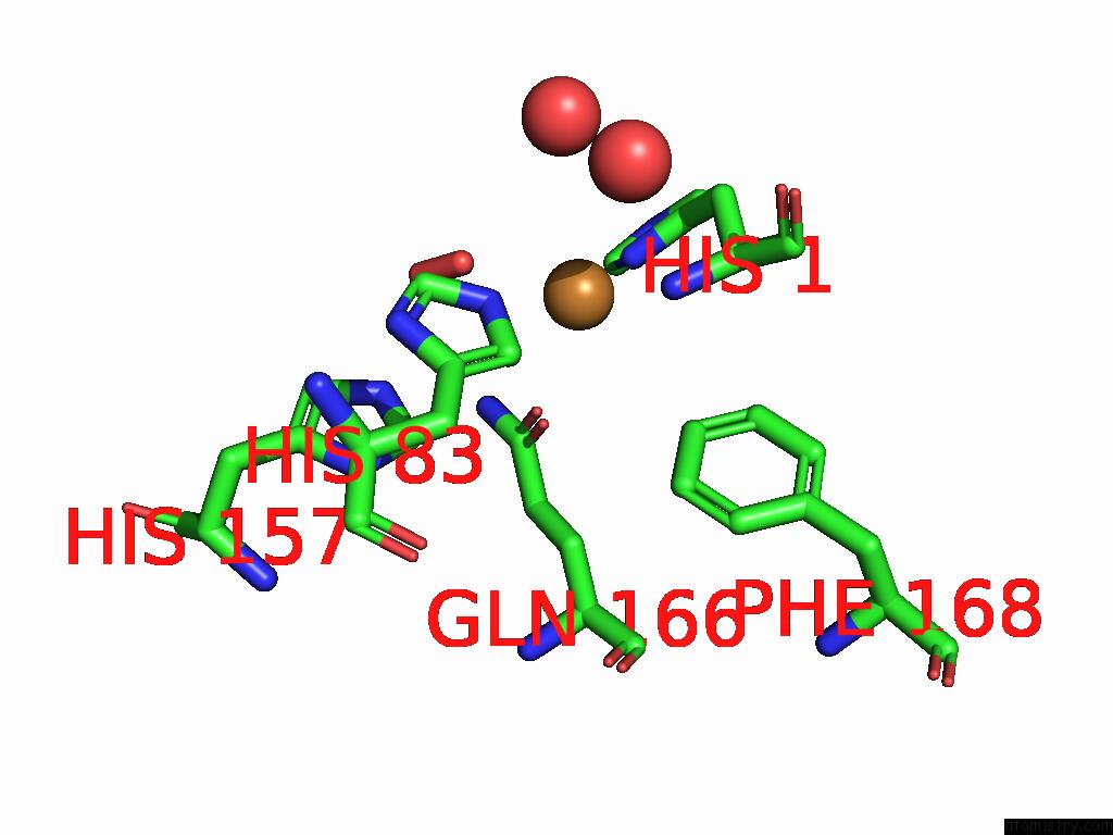
Mono view
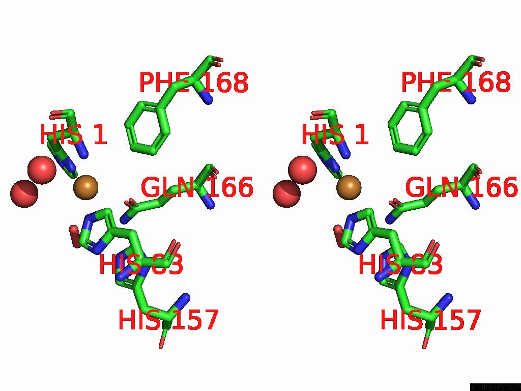
Stereo pair view
Mono view
Stereo pair view
A full contact list of Copper with other atoms in the Cu binding
site number 2 of X-Ray Crystal Structure of Y168F Variant Thermothelomyces Thermophilus Polysaccharide Monooxygenase 9E within 5.0Å range:
|
Reference:
R.I.Sayler,
W.C.Thomas,
A.J.Rose,
M.A.Marletta.
Electron Transfer in Polysaccharide Monooxygenase Catalysis Proc.Natl.Acad.Sci.Usa 2024.
ISSN: ESSN 1091-6490
DOI: 10.1073/PNAS.2411229121
Page generated: Mon Jul 14 09:44:39 2025
ISSN: ESSN 1091-6490
DOI: 10.1073/PNAS.2411229121
Last articles
Hg in 3GFHHg in 3FW0
Hg in 3FN8
Hg in 3F8E
Hg in 3FFP
Hg in 3F4X
Hg in 3F2F
Hg in 3F2H
Hg in 3F0P
Hg in 3D8D