Copper »
PDB 8bae-8gbt »
8dg0 »
Copper in PDB 8dg0: Crystal Structure of Ecdsba in A Complex with Urea
Protein crystallography data
The structure of Crystal Structure of Ecdsba in A Complex with Urea, PDB code: 8dg0
was solved by
R.L.Whitehouse,
O.V.Ilyichova,
A.J.Taylor,
with X-Ray Crystallography technique. A brief refinement statistics is given in the table below:
| Resolution Low / High (Å) | 47.46 / 2.50 |
| Space group | C 1 2 1 |
| Cell size a, b, c (Å), α, β, γ (°) | 116.341, 64.324, 75.346, 90, 125.32, 90 |
| R / Rfree (%) | 20.2 / 25.7 |
Copper Binding Sites:
The binding sites of Copper atom in the Crystal Structure of Ecdsba in A Complex with Urea
(pdb code 8dg0). This binding sites where shown within
5.0 Angstroms radius around Copper atom.
In total 2 binding sites of Copper where determined in the Crystal Structure of Ecdsba in A Complex with Urea, PDB code: 8dg0:
Jump to Copper binding site number: 1; 2;
In total 2 binding sites of Copper where determined in the Crystal Structure of Ecdsba in A Complex with Urea, PDB code: 8dg0:
Jump to Copper binding site number: 1; 2;
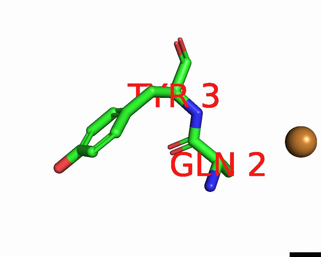
Copper binding site 1 out of 2 in 8dg0
Go back to
Copper binding site 1 out
of 2 in the Crystal Structure of Ecdsba in A Complex with Urea
Mono view

Stereo pair view
Mono view
Stereo pair view
A full contact list of Copper with other atoms in the Cu binding
site number 1 of Crystal Structure of Ecdsba in A Complex with Urea within 5.0Å range:
|
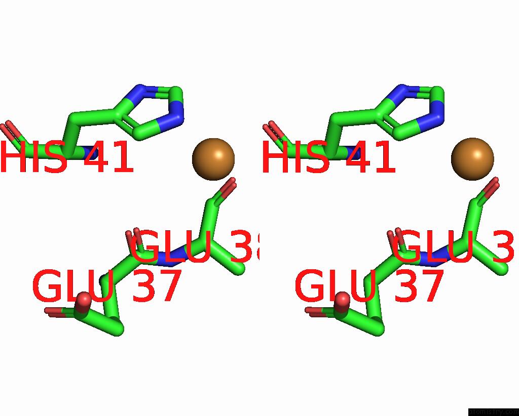
Copper binding site 2 out of 2 in 8dg0
Go back to
Copper binding site 2 out
of 2 in the Crystal Structure of Ecdsba in A Complex with Urea

Mono view
Stereo pair view
Mono view
Stereo pair view
A full contact list of Copper with other atoms in the Cu binding
site number 2 of Crystal Structure of Ecdsba in A Complex with Urea within 5.0Å range:
|
Reference:
R.L.Whitehouse,
W.S.Alwan,
O.V.Ilyichova,
A.J.Taylor,
I.R.Chandrashekaran,
B.Mohanty,
B.C.Doak,
M.J.Scanlon.
Fragment Screening Libraries For the Identification of Protein Hot Spots and Their Minimal Binding Pharmacophores Rsc Med Chem 2022.
ISSN: ESSN 2632-8682
DOI: 10.1039/D2MD00253A
Page generated: Mon Jul 14 08:53:25 2025
ISSN: ESSN 2632-8682
DOI: 10.1039/D2MD00253A
Last articles
Fe in 2YXOFe in 2YRS
Fe in 2YXC
Fe in 2YNM
Fe in 2YVJ
Fe in 2YP1
Fe in 2YU2
Fe in 2YU1
Fe in 2YQB
Fe in 2YOO