Copper »
PDB 7s1f-7xmb »
7wnp »
Copper in PDB 7wnp: Crystallographic Structure of Copper Amine Oxidase From Arthrobacter Glibiformis at Pd 7.4 Determined By Both X-Ray and Neutron Diffraction Data at 1.72 Angstrom Resolution.
Enzymatic activity of Crystallographic Structure of Copper Amine Oxidase From Arthrobacter Glibiformis at Pd 7.4 Determined By Both X-Ray and Neutron Diffraction Data at 1.72 Angstrom Resolution.
All present enzymatic activity of Crystallographic Structure of Copper Amine Oxidase From Arthrobacter Glibiformis at Pd 7.4 Determined By Both X-Ray and Neutron Diffraction Data at 1.72 Angstrom Resolution.:
1.4.3.21;
1.4.3.21;
Protein crystallography data
The structure of Crystallographic Structure of Copper Amine Oxidase From Arthrobacter Glibiformis at Pd 7.4 Determined By Both X-Ray and Neutron Diffraction Data at 1.72 Angstrom Resolution., PDB code: 7wnp
was solved by
T.Murakawa,
T.Okajima,
with X-Ray Crystallography technique. A brief refinement statistics is given in the table below:
| Resolution Low / High (Å) | N/A / 1.72 |
| Space group | C 1 2 1 |
| Cell size a, b, c (Å), α, β, γ (°) | 157.206, 61.979, 92.447, 90, 112.06, 90 |
| R / Rfree (%) | 18.9 / 23.5 |
Other elements in 7wnp:
The structure of Crystallographic Structure of Copper Amine Oxidase From Arthrobacter Glibiformis at Pd 7.4 Determined By Both X-Ray and Neutron Diffraction Data at 1.72 Angstrom Resolution. also contains other interesting chemical elements:
| Sodium | (Na) | 1 atom |
Copper Binding Sites:
The binding sites of Copper atom in the Crystallographic Structure of Copper Amine Oxidase From Arthrobacter Glibiformis at Pd 7.4 Determined By Both X-Ray and Neutron Diffraction Data at 1.72 Angstrom Resolution.
(pdb code 7wnp). This binding sites where shown within
5.0 Angstroms radius around Copper atom.
In total only one binding site of Copper was determined in the Crystallographic Structure of Copper Amine Oxidase From Arthrobacter Glibiformis at Pd 7.4 Determined By Both X-Ray and Neutron Diffraction Data at 1.72 Angstrom Resolution., PDB code: 7wnp:
In total only one binding site of Copper was determined in the Crystallographic Structure of Copper Amine Oxidase From Arthrobacter Glibiformis at Pd 7.4 Determined By Both X-Ray and Neutron Diffraction Data at 1.72 Angstrom Resolution., PDB code: 7wnp:
Copper binding site 1 out of 1 in 7wnp
Go back to
Copper binding site 1 out
of 1 in the Crystallographic Structure of Copper Amine Oxidase From Arthrobacter Glibiformis at Pd 7.4 Determined By Both X-Ray and Neutron Diffraction Data at 1.72 Angstrom Resolution.
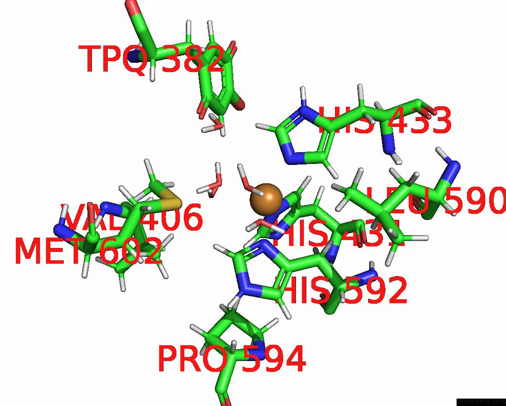
Mono view
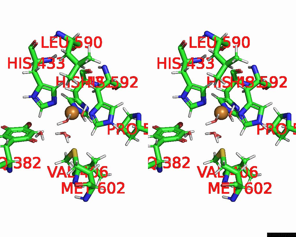
Stereo pair view
Mono view
Stereo pair view
A full contact list of Copper with other atoms in the Cu binding
site number 1 of Crystallographic Structure of Copper Amine Oxidase From Arthrobacter Glibiformis at Pd 7.4 Determined By Both X-Ray and Neutron Diffraction Data at 1.72 Angstrom Resolution. within 5.0Å range:
|
Reference:
T.Murakawa,
K.Kurihara,
M.Adachi,
K.Kusaka,
K.Tanizawa,
T.Okajima.
Re-Evaluation of Protein Neutron Crystallography with and Without X-Ray/Neutron Joint Refinement. Iucrj V. 9 342 2022.
ISSN: ESSN 2052-2525
PubMed: 35546796
DOI: 10.1107/S2052252522003657
Page generated: Mon Jul 14 08:39:37 2025
ISSN: ESSN 2052-2525
PubMed: 35546796
DOI: 10.1107/S2052252522003657
Last articles
I in 1U65I in 1THA
I in 1TUK
I in 1TF9
I in 1TB4
I in 1T6H
I in 1T6C
I in 1SD5
I in 1T4E
I in 1SO2