Copper »
PDB 7s1f-7xmb »
7vw1 »
Copper in PDB 7vw1: Structure of A Dimeric Periplasmic Protein Bound with Cuprous Ions
Protein crystallography data
The structure of Structure of A Dimeric Periplasmic Protein Bound with Cuprous Ions, PDB code: 7vw1
was solved by
J.Yang,
L.Liu,
with X-Ray Crystallography technique. A brief refinement statistics is given in the table below:
| Resolution Low / High (Å) | 34.62 / 2.49 |
| Space group | P 43 |
| Cell size a, b, c (Å), α, β, γ (°) | 77.415, 77.415, 44.933, 90, 90, 90 |
| R / Rfree (%) | 20.3 / 24.8 |
Copper Binding Sites:
The binding sites of Copper atom in the Structure of A Dimeric Periplasmic Protein Bound with Cuprous Ions
(pdb code 7vw1). This binding sites where shown within
5.0 Angstroms radius around Copper atom.
In total 5 binding sites of Copper where determined in the Structure of A Dimeric Periplasmic Protein Bound with Cuprous Ions, PDB code: 7vw1:
Jump to Copper binding site number: 1; 2; 3; 4; 5;
In total 5 binding sites of Copper where determined in the Structure of A Dimeric Periplasmic Protein Bound with Cuprous Ions, PDB code: 7vw1:
Jump to Copper binding site number: 1; 2; 3; 4; 5;
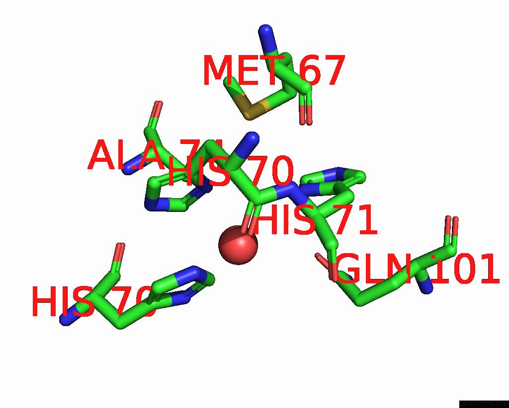
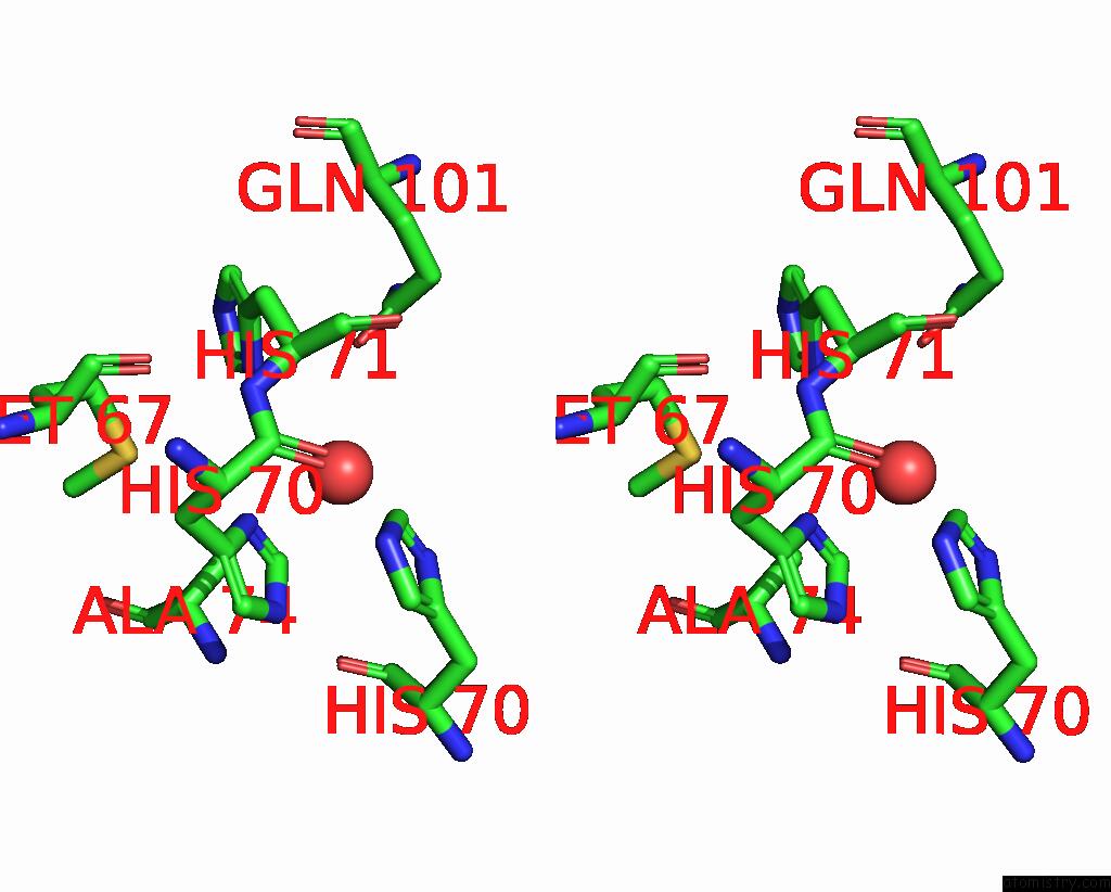
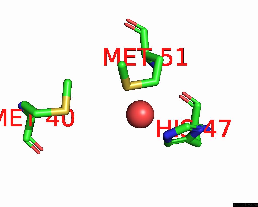
Copper binding site 1 out of 5 in 7vw1
Go back to
Copper binding site 1 out
of 5 in the Structure of A Dimeric Periplasmic Protein Bound with Cuprous Ions

Mono view
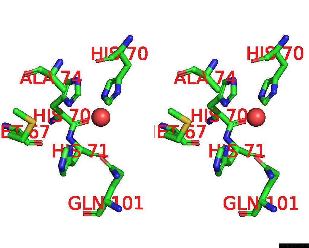
Stereo pair view
Mono view
Stereo pair view
A full contact list of Copper with other atoms in the Cu binding
site number 1 of Structure of A Dimeric Periplasmic Protein Bound with Cuprous Ions within 5.0Å range:
|
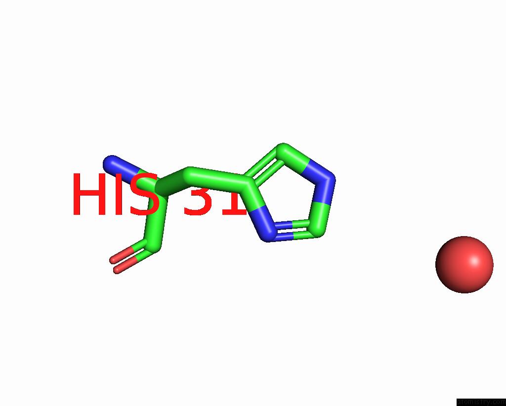
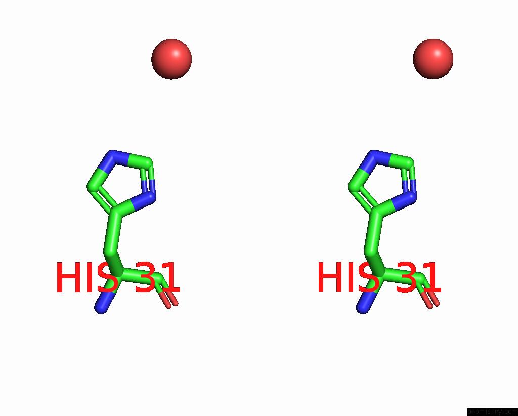
Copper binding site 2 out of 5 in 7vw1
Go back to
Copper binding site 2 out
of 5 in the Structure of A Dimeric Periplasmic Protein Bound with Cuprous Ions

Mono view
Stereo pair view
Mono view
Stereo pair view
A full contact list of Copper with other atoms in the Cu binding
site number 2 of Structure of A Dimeric Periplasmic Protein Bound with Cuprous Ions within 5.0Å range:
|
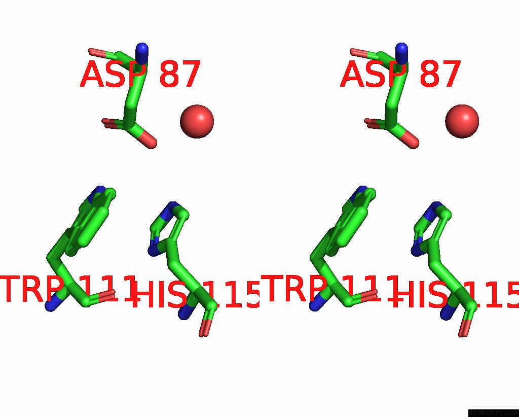
Copper binding site 3 out of 5 in 7vw1
Go back to
Copper binding site 3 out
of 5 in the Structure of A Dimeric Periplasmic Protein Bound with Cuprous Ions
Mono view
Stereo pair view
Mono view
Stereo pair view
A full contact list of Copper with other atoms in the Cu binding
site number 3 of Structure of A Dimeric Periplasmic Protein Bound with Cuprous Ions within 5.0Å range:
|
Copper binding site 4 out of 5 in 7vw1
Go back to
Copper binding site 4 out
of 5 in the Structure of A Dimeric Periplasmic Protein Bound with Cuprous Ions
Mono view

Stereo pair view
Mono view
Stereo pair view
A full contact list of Copper with other atoms in the Cu binding
site number 4 of Structure of A Dimeric Periplasmic Protein Bound with Cuprous Ions within 5.0Å range:
|
Copper binding site 5 out of 5 in 7vw1
Go back to
Copper binding site 5 out
of 5 in the Structure of A Dimeric Periplasmic Protein Bound with Cuprous Ions
Mono view
Stereo pair view
Mono view
Stereo pair view
A full contact list of Copper with other atoms in the Cu binding
site number 5 of Structure of A Dimeric Periplasmic Protein Bound with Cuprous Ions within 5.0Å range:
|
Reference:
J.Yang,
M.Gao,
J.Wang,
C.He,
X.Wang,
L.Liu.
Structural Basis of Copper Binding By A Dimeric Periplasmic Protein Forming A Six-Helical Bundle. J.Inorg.Biochem. V. 229 11728 2022.
ISSN: ISSN 0162-0134
PubMed: 35066349
DOI: 10.1016/J.JINORGBIO.2022.111728
Page generated: Mon Jul 14 08:37:41 2025
ISSN: ISSN 0162-0134
PubMed: 35066349
DOI: 10.1016/J.JINORGBIO.2022.111728
Last articles
I in 2CJ6I in 2CEO
I in 2CIW
I in 2C3N
I in 2C6C
I in 2C3V
I in 2C47
I in 2BXN
I in 2BIV
I in 2B9X