Copper »
PDB 7o3e-7pyi »
7pxm »
Copper in PDB 7pxm: X-Ray Structure of Lpmo at 1.45X10^6 Gy
Enzymatic activity of X-Ray Structure of Lpmo at 1.45X10^6 Gy
All present enzymatic activity of X-Ray Structure of Lpmo at 1.45X10^6 Gy:
1.14.99.56;
1.14.99.56;
Protein crystallography data
The structure of X-Ray Structure of Lpmo at 1.45X10^6 Gy, PDB code: 7pxm
was solved by
T.Tandrup,
L.Lo Leggio,
with X-Ray Crystallography technique. A brief refinement statistics is given in the table below:
| Resolution Low / High (Å) | 44.26 / 1.30 |
| Space group | P 41 3 2 |
| Cell size a, b, c (Å), α, β, γ (°) | 125.08, 125.08, 125.08, 90, 90, 90 |
| R / Rfree (%) | 18.5 / 20.2 |
Copper Binding Sites:
The binding sites of Copper atom in the X-Ray Structure of Lpmo at 1.45X10^6 Gy
(pdb code 7pxm). This binding sites where shown within
5.0 Angstroms radius around Copper atom.
In total only one binding site of Copper was determined in the X-Ray Structure of Lpmo at 1.45X10^6 Gy, PDB code: 7pxm:
In total only one binding site of Copper was determined in the X-Ray Structure of Lpmo at 1.45X10^6 Gy, PDB code: 7pxm:
Copper binding site 1 out of 1 in 7pxm
Go back to
Copper binding site 1 out
of 1 in the X-Ray Structure of Lpmo at 1.45X10^6 Gy
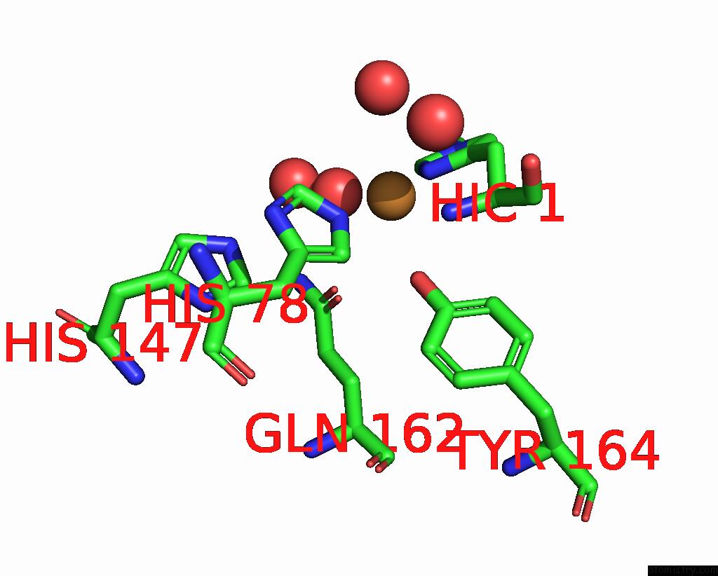
Mono view
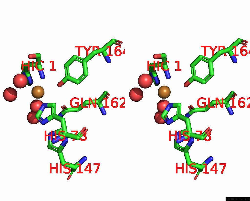
Stereo pair view
Mono view
Stereo pair view
A full contact list of Copper with other atoms in the Cu binding
site number 1 of X-Ray Structure of Lpmo at 1.45X10^6 Gy within 5.0Å range:
|
Reference:
T.Tandrup,
S.J.Muderspach,
S.Banerjee,
G.Santoni,
J.O.Ipsen,
C.Hernandez-Rollan,
M.H.H.Norholm,
K.S.Johansen,
F.Meilleur,
L.Lo Leggio.
Changes in Active-Site Geometry on X-Ray Photoreduction of A Lytic Polysaccharide Monooxygenase Active-Site Copper and Saccharide Binding. Iucrj V. 9 666 2022.
ISSN: ESSN 2052-2525
PubMed: 36071795
DOI: 10.1107/S2052252522007175
Page generated: Mon Jul 14 08:18:36 2025
ISSN: ESSN 2052-2525
PubMed: 36071795
DOI: 10.1107/S2052252522007175
Last articles
I in 2AF6I in 2ANV
I in 2A0N
I in 1XC6
I in 1ZVV
I in 1Z7J
I in 1ZME
I in 1ZCR
I in 1YVP
I in 1YRI