Copper »
PDB 5onx-5wbc »
5vg1 »
Copper in PDB 5vg1: Neutron Crystallographic Structure of A Jonesia Denitrificans Lytic Polysaccharide Monooxygenase
Enzymatic activity of Neutron Crystallographic Structure of A Jonesia Denitrificans Lytic Polysaccharide Monooxygenase
All present enzymatic activity of Neutron Crystallographic Structure of A Jonesia Denitrificans Lytic Polysaccharide Monooxygenase:
3.2.1.14;
3.2.1.14;
Copper Binding Sites:
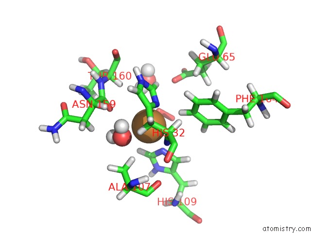
The binding sites of Copper atom in the Neutron Crystallographic Structure of A Jonesia Denitrificans Lytic Polysaccharide Monooxygenase
(pdb code 5vg1). This binding sites where shown within
5.0 Angstroms radius around Copper atom.
In total 2 binding sites of Copper where determined in the Neutron Crystallographic Structure of A Jonesia Denitrificans Lytic Polysaccharide Monooxygenase, PDB code: 5vg1:
Jump to Copper binding site number: 1; 2;
In total 2 binding sites of Copper where determined in the Neutron Crystallographic Structure of A Jonesia Denitrificans Lytic Polysaccharide Monooxygenase, PDB code: 5vg1:
Jump to Copper binding site number: 1; 2;
Copper binding site 1 out of 2 in 5vg1
Go back to
Copper binding site 1 out
of 2 in the Neutron Crystallographic Structure of A Jonesia Denitrificans Lytic Polysaccharide Monooxygenase

Mono view
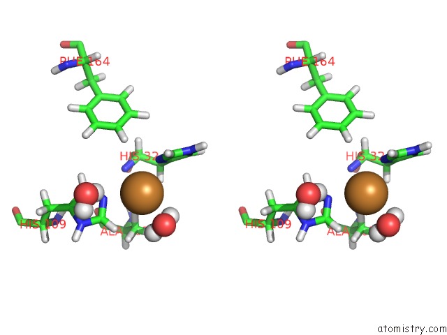
Stereo pair view
Mono view
Stereo pair view
A full contact list of Copper with other atoms in the Cu binding
site number 1 of Neutron Crystallographic Structure of A Jonesia Denitrificans Lytic Polysaccharide Monooxygenase within 5.0Å range:
|
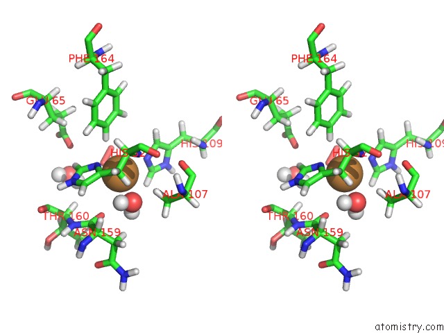
Copper binding site 2 out of 2 in 5vg1
Go back to
Copper binding site 2 out
of 2 in the Neutron Crystallographic Structure of A Jonesia Denitrificans Lytic Polysaccharide Monooxygenase
Mono view
Stereo pair view
Mono view
Stereo pair view
A full contact list of Copper with other atoms in the Cu binding
site number 2 of Neutron Crystallographic Structure of A Jonesia Denitrificans Lytic Polysaccharide Monooxygenase within 5.0Å range:
|
Reference:
J.P.Bacik,
S.Mekasha,
Z.Forsberg,
A.Y.Kovalevsky,
G.Vaaje-Kolstad,
V.G.H.Eijsink,
J.C.Nix,
L.Coates,
M.J.Cuneo,
C.J.Unkefer,
J.C.Chen.
Neutron and Atomic Resolution X-Ray Structures of A Lytic Polysaccharide Monooxygenase Reveal Copper-Mediated Dioxygen Binding and Evidence For N-Terminal Deprotonation. Biochemistry V. 56 2529 2017.
ISSN: ISSN 1520-4995
PubMed: 28481095
DOI: 10.1021/ACS.BIOCHEM.7B00019
Page generated: Mon Jul 14 05:29:10 2025
ISSN: ISSN 1520-4995
PubMed: 28481095
DOI: 10.1021/ACS.BIOCHEM.7B00019
Last articles
Hg in 1G52Hg in 1G53
Hg in 1G54
Hg in 1G48
Hg in 1G4J
Hg in 1G4O
Hg in 1FMJ
Hg in 1G46
Hg in 1G45
Hg in 1G3Z