Copper »
PDB 4ysu-5c92 »
5b7f »
Copper in PDB 5b7f: Structure of Cueo - the Signal Peptide Was Truncated By HRV3C Protease
Protein crystallography data
The structure of Structure of Cueo - the Signal Peptide Was Truncated By HRV3C Protease, PDB code: 5b7f
was solved by
M.Akter,
Y.Higuchi,
N.Shibata,
with X-Ray Crystallography technique. A brief refinement statistics is given in the table below:
| Resolution Low / High (Å) | 35.11 / 1.45 |
| Space group | P 1 21 1 |
| Cell size a, b, c (Å), α, β, γ (°) | 49.513, 88.786, 53.951, 90.00, 94.24, 90.00 |
| R / Rfree (%) | 15.3 / 18.1 |
Other elements in 5b7f:
The structure of Structure of Cueo - the Signal Peptide Was Truncated By HRV3C Protease also contains other interesting chemical elements:
| Calcium | (Ca) | 1 atom |
Copper Binding Sites:
The binding sites of Copper atom in the Structure of Cueo - the Signal Peptide Was Truncated By HRV3C Protease
(pdb code 5b7f). This binding sites where shown within
5.0 Angstroms radius around Copper atom.
In total 4 binding sites of Copper where determined in the Structure of Cueo - the Signal Peptide Was Truncated By HRV3C Protease, PDB code: 5b7f:
Jump to Copper binding site number: 1; 2; 3; 4;
In total 4 binding sites of Copper where determined in the Structure of Cueo - the Signal Peptide Was Truncated By HRV3C Protease, PDB code: 5b7f:
Jump to Copper binding site number: 1; 2; 3; 4;
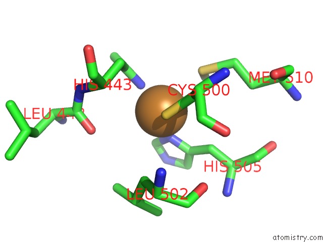
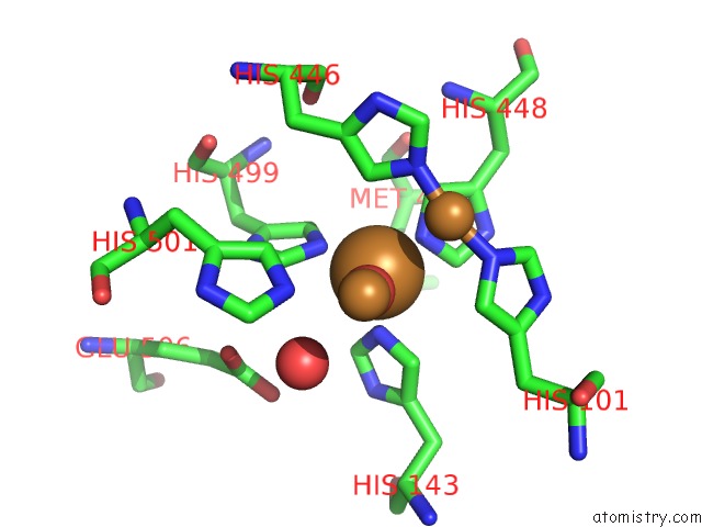
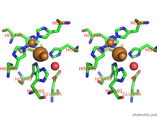
Copper binding site 1 out of 4 in 5b7f
Go back to
Copper binding site 1 out
of 4 in the Structure of Cueo - the Signal Peptide Was Truncated By HRV3C Protease
Mono view
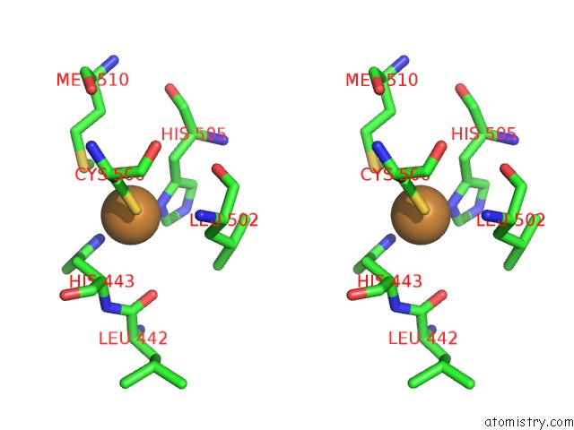
Stereo pair view
Mono view
Stereo pair view
A full contact list of Copper with other atoms in the Cu binding
site number 1 of Structure of Cueo - the Signal Peptide Was Truncated By HRV3C Protease within 5.0Å range:
|
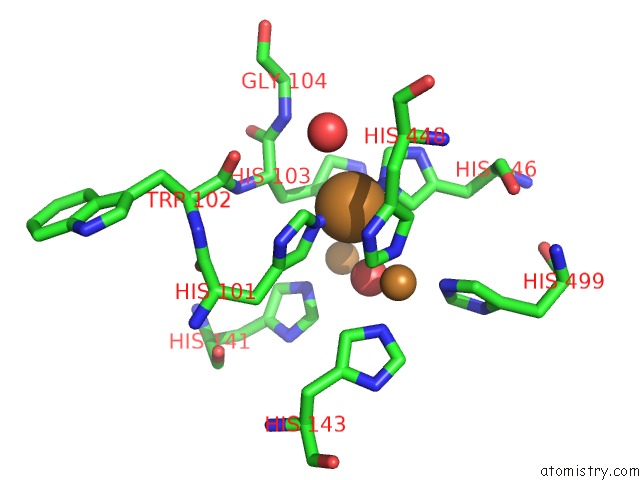
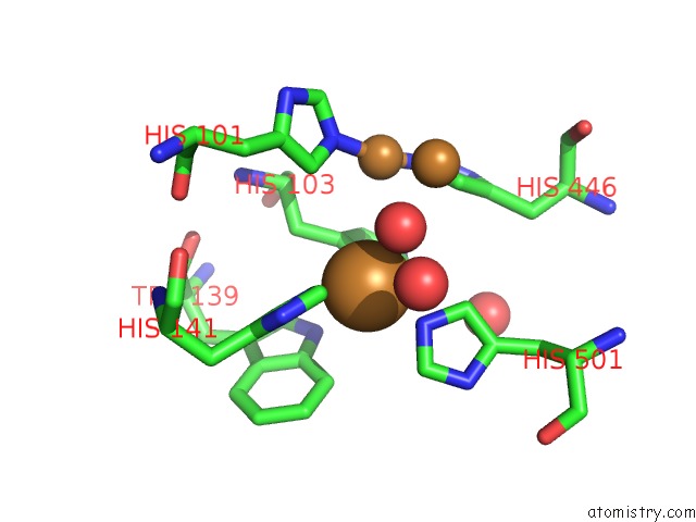
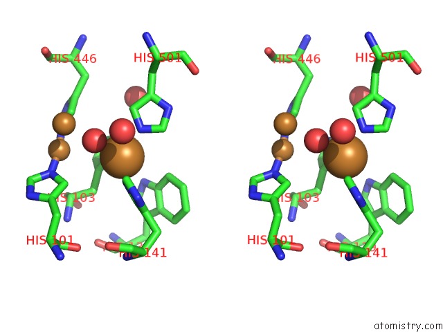
Copper binding site 2 out of 4 in 5b7f
Go back to
Copper binding site 2 out
of 4 in the Structure of Cueo - the Signal Peptide Was Truncated By HRV3C Protease
Mono view
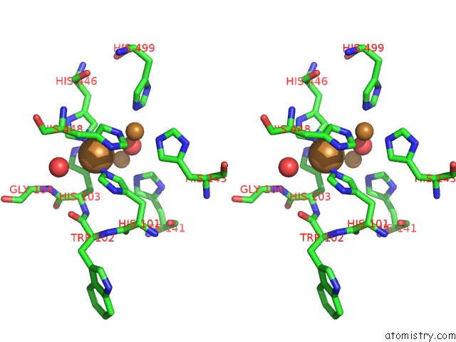
Stereo pair view
Mono view
Stereo pair view
A full contact list of Copper with other atoms in the Cu binding
site number 2 of Structure of Cueo - the Signal Peptide Was Truncated By HRV3C Protease within 5.0Å range:
|
Copper binding site 3 out of 4 in 5b7f
Go back to
Copper binding site 3 out
of 4 in the Structure of Cueo - the Signal Peptide Was Truncated By HRV3C Protease
Mono view
Stereo pair view
Mono view
Stereo pair view
A full contact list of Copper with other atoms in the Cu binding
site number 3 of Structure of Cueo - the Signal Peptide Was Truncated By HRV3C Protease within 5.0Å range:
|
Copper binding site 4 out of 4 in 5b7f
Go back to
Copper binding site 4 out
of 4 in the Structure of Cueo - the Signal Peptide Was Truncated By HRV3C Protease
Mono view
Stereo pair view
Mono view
Stereo pair view
A full contact list of Copper with other atoms in the Cu binding
site number 4 of Structure of Cueo - the Signal Peptide Was Truncated By HRV3C Protease within 5.0Å range:
|
Reference:
M.Akter,
C.Inoue,
H.Komori,
N.Matsuda,
T.Sakurai,
K.Kataoka,
Y.Higuchi,
N.Shibata.
Biochemical, Spectroscopic and X-Ray Structural Analysis of Deuterated Multicopper Oxidase Cueo Prepared From A New Expression Construct For Neutron Crystallography Acta Crystallogr.,Sect.F V. 72 788 2016.
ISSN: ESSN 2053-230X
PubMed: 27710945
DOI: 10.1107/S2053230X1601400X
Page generated: Mon Jul 14 04:27:30 2025
ISSN: ESSN 2053-230X
PubMed: 27710945
DOI: 10.1107/S2053230X1601400X
Last articles
Mg in 4DV3Mg in 4DV2
Mg in 4DV0
Mg in 4DV1
Mg in 4DUZ
Mg in 4DUY
Mg in 4DR7
Mg in 4DR6
Mg in 4DR5
Mg in 4DUX