Copper »
PDB 4ysu-5c92 »
4yzw »
Copper in PDB 4yzw: Crystal Structure of AGPPO8
Protein crystallography data
The structure of Crystal Structure of AGPPO8, PDB code: 4yzw
was solved by
Y.Hu,
with X-Ray Crystallography technique. A brief refinement statistics is given in the table below:
| Resolution Low / High (Å) | 44.32 / 2.60 |
| Space group | P 1 21 1 |
| Cell size a, b, c (Å), α, β, γ (°) | 75.583, 106.583, 92.110, 90.00, 105.79, 90.00 |
| R / Rfree (%) | 18.8 / 23.3 |
Copper Binding Sites:
The binding sites of Copper atom in the Crystal Structure of AGPPO8
(pdb code 4yzw). This binding sites where shown within
5.0 Angstroms radius around Copper atom.
In total 4 binding sites of Copper where determined in the Crystal Structure of AGPPO8, PDB code: 4yzw:
Jump to Copper binding site number: 1; 2; 3; 4;
In total 4 binding sites of Copper where determined in the Crystal Structure of AGPPO8, PDB code: 4yzw:
Jump to Copper binding site number: 1; 2; 3; 4;
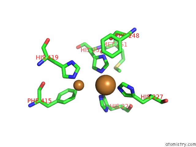
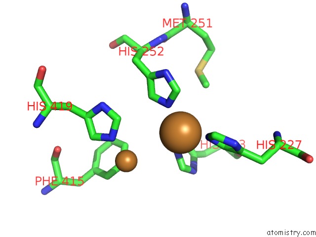
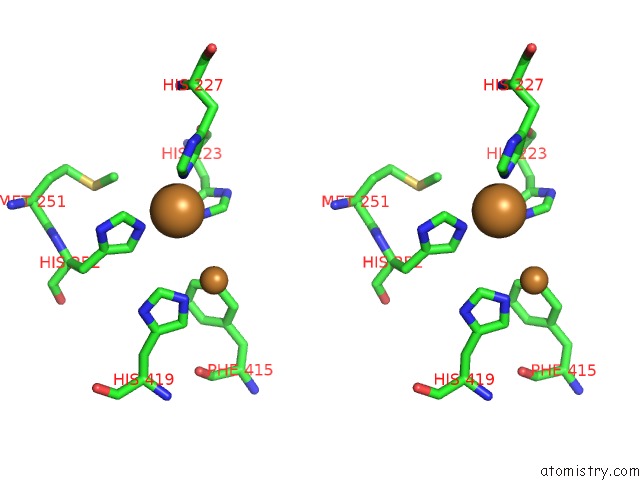
Copper binding site 1 out of 4 in 4yzw
Go back to
Copper binding site 1 out
of 4 in the Crystal Structure of AGPPO8
Mono view
Stereo pair view
Mono view
Stereo pair view
A full contact list of Copper with other atoms in the Cu binding
site number 1 of Crystal Structure of AGPPO8 within 5.0Å range:
|
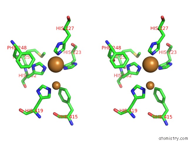
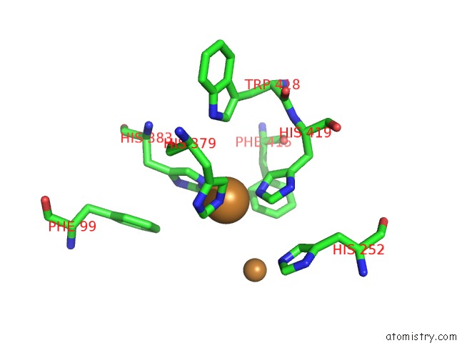
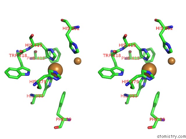
Copper binding site 2 out of 4 in 4yzw
Go back to
Copper binding site 2 out
of 4 in the Crystal Structure of AGPPO8
Mono view
Stereo pair view
Mono view
Stereo pair view
A full contact list of Copper with other atoms in the Cu binding
site number 2 of Crystal Structure of AGPPO8 within 5.0Å range:
|
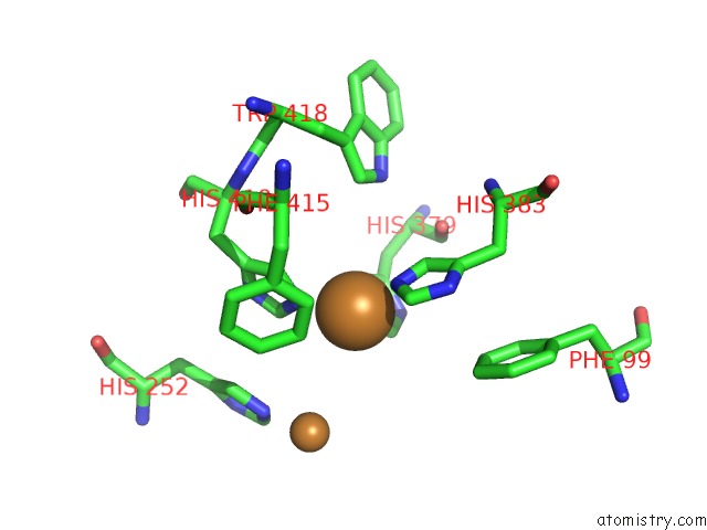
Copper binding site 3 out of 4 in 4yzw
Go back to
Copper binding site 3 out
of 4 in the Crystal Structure of AGPPO8
Mono view
Stereo pair view
Mono view
Stereo pair view
A full contact list of Copper with other atoms in the Cu binding
site number 3 of Crystal Structure of AGPPO8 within 5.0Å range:
|
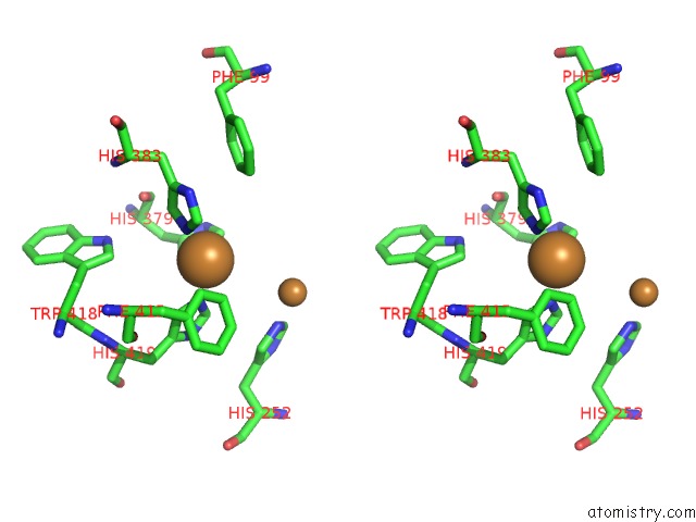
Copper binding site 4 out of 4 in 4yzw
Go back to
Copper binding site 4 out
of 4 in the Crystal Structure of AGPPO8
Mono view
Stereo pair view
Mono view
Stereo pair view
A full contact list of Copper with other atoms in the Cu binding
site number 4 of Crystal Structure of AGPPO8 within 5.0Å range:
|
Reference:
Y.Hu,
Y.Wang,
J.Deng,
H.Jiang.
The Structure of A Prophenoloxidase (Ppo) From Anopheles Gambiae Provides New Insights Into the Mechanism of Ppo Activation. Bmc Biol. V. 14 2 2016.
ISSN: ESSN 1741-7007
PubMed: 26732497
DOI: 10.1186/S12915-015-0225-2
Page generated: Mon Jul 14 04:15:03 2025
ISSN: ESSN 1741-7007
PubMed: 26732497
DOI: 10.1186/S12915-015-0225-2
Last articles
Mg in 4DV3Mg in 4DV2
Mg in 4DV0
Mg in 4DV1
Mg in 4DUZ
Mg in 4DUY
Mg in 4DR7
Mg in 4DR6
Mg in 4DR5
Mg in 4DUX