Copper »
PDB 4mfh-4rkn »
4oak »
Copper in PDB 4oak: Crystal Structure of Vancomycin Resistance D,D-Dipeptidase/D,D- Pentapeptidase Vanxyc D59S Mutant in Complex with D-Alanine-D-Alanine and Copper (II)
Protein crystallography data
The structure of Crystal Structure of Vancomycin Resistance D,D-Dipeptidase/D,D- Pentapeptidase Vanxyc D59S Mutant in Complex with D-Alanine-D-Alanine and Copper (II), PDB code: 4oak
was solved by
P.J.Stogios,
E.Evdokimova,
D.Meziane-Cherif,
R.Di Leo,
V.Yim,
P.Courvalin,
A.Savchenko,
W.F.Anderson,
Center For Structural Genomics Ofinfectious Diseases (Csgid),
with X-Ray Crystallography technique. A brief refinement statistics is given in the table below:
| Resolution Low / High (Å) | 19.33 / 2.00 |
| Space group | P 1 |
| Cell size a, b, c (Å), α, β, γ (°) | 41.939, 43.083, 66.316, 80.53, 74.52, 64.01 |
| R / Rfree (%) | 18.9 / 23.5 |
Other elements in 4oak:
The structure of Crystal Structure of Vancomycin Resistance D,D-Dipeptidase/D,D- Pentapeptidase Vanxyc D59S Mutant in Complex with D-Alanine-D-Alanine and Copper (II) also contains other interesting chemical elements:
| Magnesium | (Mg) | 1 atom |
| Chlorine | (Cl) | 6 atoms |
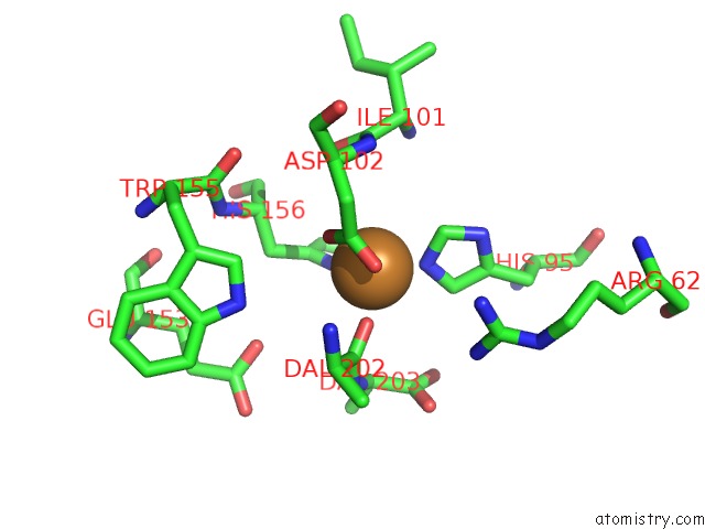
Copper Binding Sites:
The binding sites of Copper atom in the Crystal Structure of Vancomycin Resistance D,D-Dipeptidase/D,D- Pentapeptidase Vanxyc D59S Mutant in Complex with D-Alanine-D-Alanine and Copper (II)
(pdb code 4oak). This binding sites where shown within
5.0 Angstroms radius around Copper atom.
In total 2 binding sites of Copper where determined in the Crystal Structure of Vancomycin Resistance D,D-Dipeptidase/D,D- Pentapeptidase Vanxyc D59S Mutant in Complex with D-Alanine-D-Alanine and Copper (II), PDB code: 4oak:
Jump to Copper binding site number: 1; 2;
In total 2 binding sites of Copper where determined in the Crystal Structure of Vancomycin Resistance D,D-Dipeptidase/D,D- Pentapeptidase Vanxyc D59S Mutant in Complex with D-Alanine-D-Alanine and Copper (II), PDB code: 4oak:
Jump to Copper binding site number: 1; 2;
Copper binding site 1 out of 2 in 4oak
Go back to
Copper binding site 1 out
of 2 in the Crystal Structure of Vancomycin Resistance D,D-Dipeptidase/D,D- Pentapeptidase Vanxyc D59S Mutant in Complex with D-Alanine-D-Alanine and Copper (II)

Mono view

Stereo pair view
Mono view
Stereo pair view
A full contact list of Copper with other atoms in the Cu binding
site number 1 of Crystal Structure of Vancomycin Resistance D,D-Dipeptidase/D,D- Pentapeptidase Vanxyc D59S Mutant in Complex with D-Alanine-D-Alanine and Copper (II) within 5.0Å range:
|
Copper binding site 2 out of 2 in 4oak
Go back to
Copper binding site 2 out
of 2 in the Crystal Structure of Vancomycin Resistance D,D-Dipeptidase/D,D- Pentapeptidase Vanxyc D59S Mutant in Complex with D-Alanine-D-Alanine and Copper (II)
Mono view

Stereo pair view
Mono view
Stereo pair view
A full contact list of Copper with other atoms in the Cu binding
site number 2 of Crystal Structure of Vancomycin Resistance D,D-Dipeptidase/D,D- Pentapeptidase Vanxyc D59S Mutant in Complex with D-Alanine-D-Alanine and Copper (II) within 5.0Å range:
|
Reference:
D.Meziane-Cherif,
P.J.Stogios,
E.Evdokimova,
A.Savchenko,
P.Courvalin.
Structural Basis For the Evolution of Vancomycin Resistance D,D-Peptidases. Proc.Natl.Acad.Sci.Usa V. 111 5872 2014.
ISSN: ISSN 0027-8424
PubMed: 24711382
DOI: 10.1073/PNAS.1402259111
Page generated: Mon Jul 14 03:57:55 2025
ISSN: ISSN 0027-8424
PubMed: 24711382
DOI: 10.1073/PNAS.1402259111
Last articles
Mg in 2WOQMg in 2WOJ
Mg in 2WQS
Mg in 2WQN
Mg in 2WOG
Mg in 2WNL
Mg in 2WLL
Mg in 2WNH
Mg in 2WMB
Mg in 2WNA