Copper »
PDB 4mfh-4rkn »
4o65 »
Copper in PDB 4o65: Crystal Structure of the Cupredoxin Domain of Amob From Nitrosocaldus Yellowstonii
Protein crystallography data
The structure of Crystal Structure of the Cupredoxin Domain of Amob From Nitrosocaldus Yellowstonii, PDB code: 4o65
was solved by
T.J.Lawton,
J.Ham,
T.Sun,
A.C.Rosenzweig,
with X-Ray Crystallography technique. A brief refinement statistics is given in the table below:
| Resolution Low / High (Å) | 22.43 / 1.80 |
| Space group | P 43 21 2 |
| Cell size a, b, c (Å), α, β, γ (°) | 47.886, 47.886, 192.614, 90.00, 90.00, 90.00 |
| R / Rfree (%) | 18.1 / 22.9 |
Copper Binding Sites:
The binding sites of Copper atom in the Crystal Structure of the Cupredoxin Domain of Amob From Nitrosocaldus Yellowstonii
(pdb code 4o65). This binding sites where shown within
5.0 Angstroms radius around Copper atom.
In total only one binding site of Copper was determined in the Crystal Structure of the Cupredoxin Domain of Amob From Nitrosocaldus Yellowstonii, PDB code: 4o65:
In total only one binding site of Copper was determined in the Crystal Structure of the Cupredoxin Domain of Amob From Nitrosocaldus Yellowstonii, PDB code: 4o65:
Copper binding site 1 out of 1 in 4o65
Go back to
Copper binding site 1 out
of 1 in the Crystal Structure of the Cupredoxin Domain of Amob From Nitrosocaldus Yellowstonii
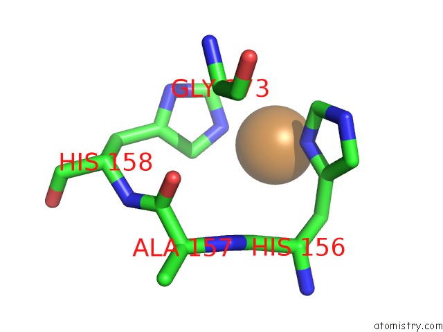
Mono view
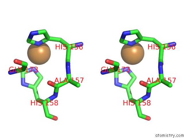
Stereo pair view
Mono view
Stereo pair view
A full contact list of Copper with other atoms in the Cu binding
site number 1 of Crystal Structure of the Cupredoxin Domain of Amob From Nitrosocaldus Yellowstonii within 5.0Å range:
|
Reference:
T.J.Lawton,
J.Ham,
T.Sun,
A.C.Rosenzweig.
Structural Conservation of the B Subunit in the Ammonia Monooxygenase/Particulate Methane Monooxygenase Superfamily. Proteins V. 82 2263 2014.
ISSN: ISSN 0887-3585
PubMed: 24523098
DOI: 10.1002/PROT.24535
Page generated: Mon Jul 14 03:57:48 2025
ISSN: ISSN 0887-3585
PubMed: 24523098
DOI: 10.1002/PROT.24535
Last articles
Mn in 9LJUMn in 9LJW
Mn in 9LJS
Mn in 9LJR
Mn in 9LJT
Mn in 9LJV
Mg in 9UA2
Mg in 9R96
Mg in 9VM1
Mg in 9P01