Copper »
PDB 3x2q-4b5q »
4alr »
Copper in PDB 4alr: X-Ray Photoreduction of Polysaccharide Monooxygenase CBM33
Protein crystallography data
The structure of X-Ray Photoreduction of Polysaccharide Monooxygenase CBM33, PDB code: 4alr
was solved by
M.Gudmundsson,
M.Wu,
T.Ishida,
M.H.Momeni,
G.Vaaje-Kolstad,
V.Eijsink,
M.Sandgren,
with X-Ray Crystallography technique. A brief refinement statistics is given in the table below:
| Resolution Low / High (Å) | 39.62 / 1.49 |
| Space group | P 21 21 21 |
| Cell size a, b, c (Å), α, β, γ (°) | 43.420, 48.578, 68.455, 90.00, 90.00, 90.00 |
| R / Rfree (%) | 16.1 / 18.8 |
Copper Binding Sites:
The binding sites of Copper atom in the X-Ray Photoreduction of Polysaccharide Monooxygenase CBM33
(pdb code 4alr). This binding sites where shown within
5.0 Angstroms radius around Copper atom.
In total only one binding site of Copper was determined in the X-Ray Photoreduction of Polysaccharide Monooxygenase CBM33, PDB code: 4alr:
In total only one binding site of Copper was determined in the X-Ray Photoreduction of Polysaccharide Monooxygenase CBM33, PDB code: 4alr:
Copper binding site 1 out of 1 in 4alr
Go back to
Copper binding site 1 out
of 1 in the X-Ray Photoreduction of Polysaccharide Monooxygenase CBM33
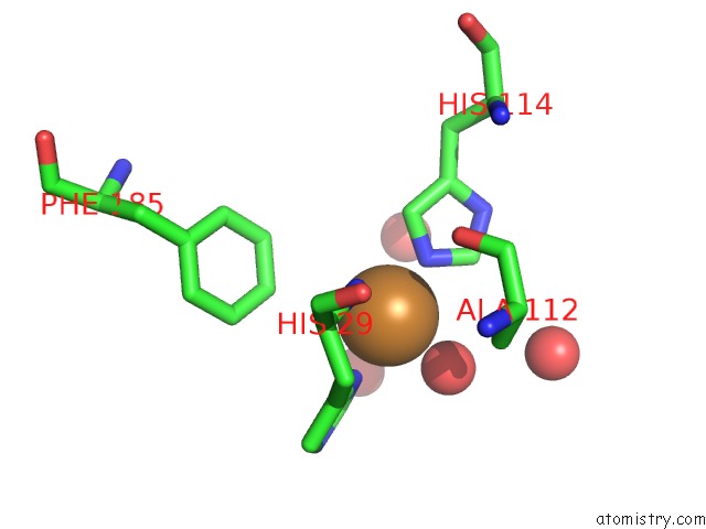
Mono view
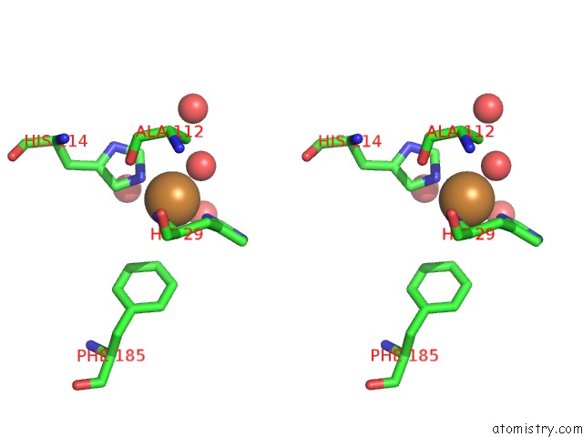
Stereo pair view
Mono view
Stereo pair view
A full contact list of Copper with other atoms in the Cu binding
site number 1 of X-Ray Photoreduction of Polysaccharide Monooxygenase CBM33 within 5.0Å range:
|
Reference:
M.Gudmundsson,
S.Kim,
M.Wu,
T.Ishida,
M.Haddad Momeni,
G.Vaaje-Kolstad,
D.Lundberg,
A.Royant,
J.Stahlberg,
V.G.Eijsink,
G.T.Beckham,
M.Sandgren.
Structural and Electronic Snapshots During the Transition From A Cu(II) to Cu(I) Metal Center of A Lytic Polysaccharide Monooxygenase By X-Ray Photo-Reduction. J.Biol.Chem. V. 289 18782 2014.
ISSN: ISSN 0021-9258
PubMed: 24828494
DOI: 10.1074/JBC.M114.563494
Page generated: Mon Jul 14 03:24:10 2025
ISSN: ISSN 0021-9258
PubMed: 24828494
DOI: 10.1074/JBC.M114.563494
Last articles
Hg in 1PIMHg in 1PFR
Hg in 1P5S
Hg in 1OPE
Hg in 1PF5
Hg in 1OF5
Hg in 1OKM
Hg in 1OKL
Hg in 1OKN
Hg in 1NAQ