Copper »
PDB 2pp8-2vr6 »
2tir »
Copper in PDB 2tir: Crystal Structure Analysis of A Mutant Escherichia Coli Thioredoxin in Which Lysine 36 Is Replaced By Glutamic Acid
Protein crystallography data
The structure of Crystal Structure Analysis of A Mutant Escherichia Coli Thioredoxin in Which Lysine 36 Is Replaced By Glutamic Acid, PDB code: 2tir
was solved by
M.Nikkola,
F.K.Gleason,
J.A.Fuchs,
H.Eklund,
with X-Ray Crystallography technique. A brief refinement statistics is given in the table below:
| Resolution Low / High (Å) | N/A / 2.00 |
| Space group | P 21 21 21 |
| Cell size a, b, c (Å), α, β, γ (°) | 50.800, 26.800, 80.700, 90.00, 90.00, 90.00 |
| R / Rfree (%) | 19.9 / n/a |
Copper Binding Sites:
The binding sites of Copper atom in the Crystal Structure Analysis of A Mutant Escherichia Coli Thioredoxin in Which Lysine 36 Is Replaced By Glutamic Acid
(pdb code 2tir). This binding sites where shown within
5.0 Angstroms radius around Copper atom.
In total only one binding site of Copper was determined in the Crystal Structure Analysis of A Mutant Escherichia Coli Thioredoxin in Which Lysine 36 Is Replaced By Glutamic Acid, PDB code: 2tir:
In total only one binding site of Copper was determined in the Crystal Structure Analysis of A Mutant Escherichia Coli Thioredoxin in Which Lysine 36 Is Replaced By Glutamic Acid, PDB code: 2tir:
Copper binding site 1 out of 1 in 2tir
Go back to
Copper binding site 1 out
of 1 in the Crystal Structure Analysis of A Mutant Escherichia Coli Thioredoxin in Which Lysine 36 Is Replaced By Glutamic Acid
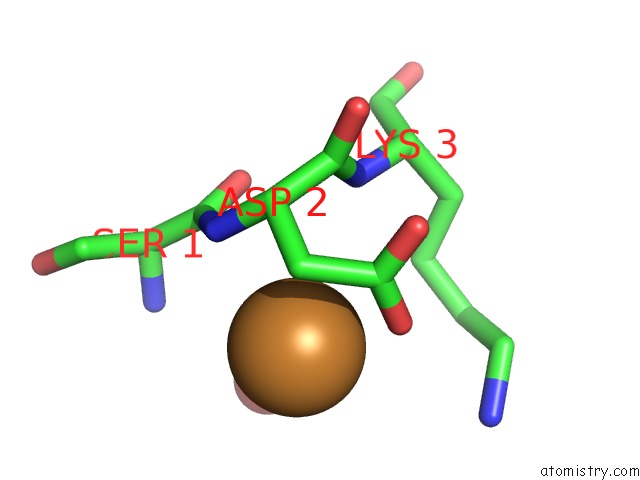
Mono view
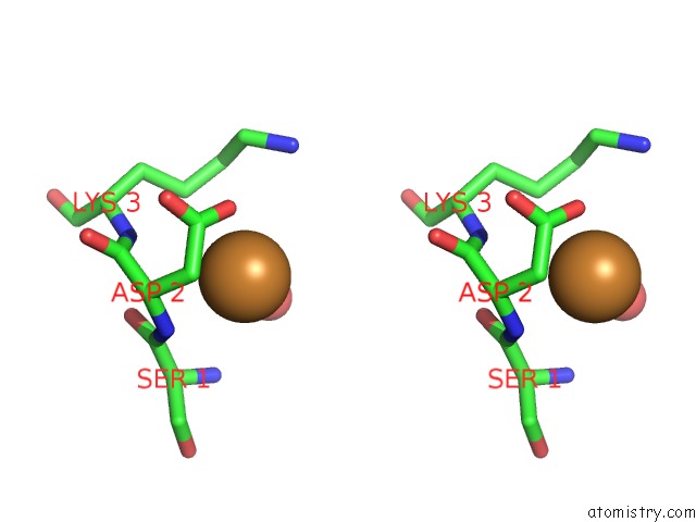
Stereo pair view
Mono view
Stereo pair view
A full contact list of Copper with other atoms in the Cu binding
site number 1 of Crystal Structure Analysis of A Mutant Escherichia Coli Thioredoxin in Which Lysine 36 Is Replaced By Glutamic Acid within 5.0Å range:
|
Reference:
M.Nikkola,
F.K.Gleason,
J.A.Fuchs,
H.Eklund.
Crystal Structure Analysis of A Mutant Escherichia Coli Thioredoxin in Which Lysine 36 Is Replaced By Glutamic Acid. Biochemistry V. 32 5093 1993.
ISSN: ISSN 0006-2960
PubMed: 8098620
DOI: 10.1021/BI00070A017
Page generated: Wed Jul 31 00:02:13 2024
ISSN: ISSN 0006-2960
PubMed: 8098620
DOI: 10.1021/BI00070A017
Last articles
Zn in 9MJ5Zn in 9HNW
Zn in 9G0L
Zn in 9FNE
Zn in 9DZN
Zn in 9E0I
Zn in 9D32
Zn in 9DAK
Zn in 8ZXC
Zn in 8ZUF