Copper »
PDB 2fqd-2idf »
2gim »
Copper in PDB 2gim: 1.6 Angstrom Structure of Plastocyanin From Anabaena Variabilis
Protein crystallography data
The structure of 1.6 Angstrom Structure of Plastocyanin From Anabaena Variabilis, PDB code: 2gim
was solved by
L.Schmidt,
P.Harris,
H.E.M.Christensen,
with X-Ray Crystallography technique. A brief refinement statistics is given in the table below:
| Resolution Low / High (Å) | 19.19 / 1.60 |
| Space group | P 21 21 21 |
| Cell size a, b, c (Å), α, β, γ (°) | 67.850, 45.810, 63.410, 90.00, 90.00, 90.00 |
| R / Rfree (%) | 20.9 / 26 |
Copper Binding Sites:
The binding sites of Copper atom in the 1.6 Angstrom Structure of Plastocyanin From Anabaena Variabilis
(pdb code 2gim). This binding sites where shown within
5.0 Angstroms radius around Copper atom.
In total 2 binding sites of Copper where determined in the 1.6 Angstrom Structure of Plastocyanin From Anabaena Variabilis, PDB code: 2gim:
Jump to Copper binding site number: 1; 2;
In total 2 binding sites of Copper where determined in the 1.6 Angstrom Structure of Plastocyanin From Anabaena Variabilis, PDB code: 2gim:
Jump to Copper binding site number: 1; 2;
Copper binding site 1 out of 2 in 2gim
Go back to
Copper binding site 1 out
of 2 in the 1.6 Angstrom Structure of Plastocyanin From Anabaena Variabilis
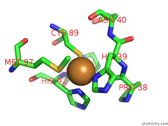
Mono view
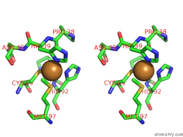
Stereo pair view
Mono view
Stereo pair view
A full contact list of Copper with other atoms in the Cu binding
site number 1 of 1.6 Angstrom Structure of Plastocyanin From Anabaena Variabilis within 5.0Å range:
|
Copper binding site 2 out of 2 in 2gim
Go back to
Copper binding site 2 out
of 2 in the 1.6 Angstrom Structure of Plastocyanin From Anabaena Variabilis
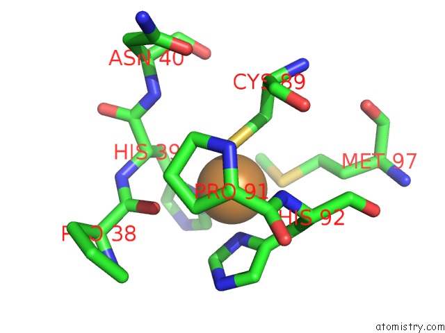
Mono view
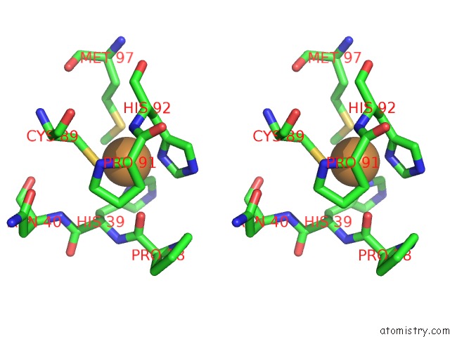
Stereo pair view
Mono view
Stereo pair view
A full contact list of Copper with other atoms in the Cu binding
site number 2 of 1.6 Angstrom Structure of Plastocyanin From Anabaena Variabilis within 5.0Å range:
|
Reference:
L.Schmidt,
H.E.Christensen,
P.Harris.
Structure of Plastocyanin From the Cyanobacterium Anabaena Variabilis. Acta Crystallogr.,Sect.D V. 62 1022 2006.
ISSN: ISSN 0907-4449
PubMed: 16929103
DOI: 10.1107/S0907444906023638
Page generated: Mon Jul 14 01:05:37 2025
ISSN: ISSN 0907-4449
PubMed: 16929103
DOI: 10.1107/S0907444906023638
Last articles
I in 7WYJI in 7YWW
I in 8A24
I in 7Z76
I in 7YDX
I in 7XME
I in 7YUZ
I in 7XMK
I in 7XC1
I in 7WWN