Copper »
PDB 1oe2-1rjp »
1rcy »
Copper in PDB 1rcy: Rusticyanin (Rc) From Thiobacillus Ferrooxidans
Protein crystallography data
The structure of Rusticyanin (Rc) From Thiobacillus Ferrooxidans, PDB code: 1rcy
was solved by
R.L.Walter,
A.M.Friedman,
S.E.Ealick,
R.C.Blake Ii,
P.Proctor,
M.Shoham,
with X-Ray Crystallography technique. A brief refinement statistics is given in the table below:
| Resolution Low / High (Å) | 10.00 / 1.90 |
| Space group | P 1 21 1 |
| Cell size a, b, c (Å), α, β, γ (°) | 32.510, 60.670, 38.140, 90.00, 108.42, 90.00 |
| R / Rfree (%) | 17.5 / 25.2 |
Copper Binding Sites:
The binding sites of Copper atom in the Rusticyanin (Rc) From Thiobacillus Ferrooxidans
(pdb code 1rcy). This binding sites where shown within
5.0 Angstroms radius around Copper atom.
In total only one binding site of Copper was determined in the Rusticyanin (Rc) From Thiobacillus Ferrooxidans, PDB code: 1rcy:
In total only one binding site of Copper was determined in the Rusticyanin (Rc) From Thiobacillus Ferrooxidans, PDB code: 1rcy:
Copper binding site 1 out of 1 in 1rcy
Go back to
Copper binding site 1 out
of 1 in the Rusticyanin (Rc) From Thiobacillus Ferrooxidans
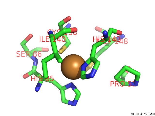
Mono view
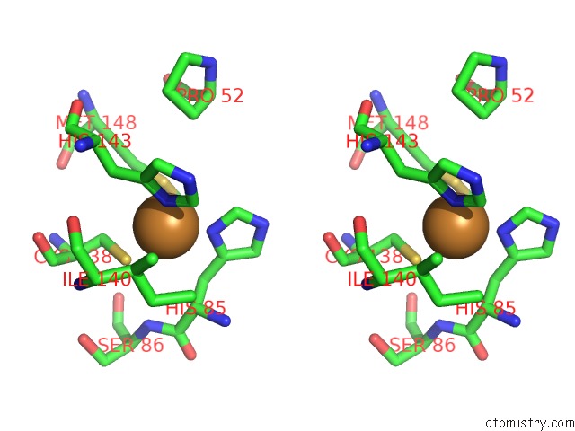
Stereo pair view
Mono view
Stereo pair view
A full contact list of Copper with other atoms in the Cu binding
site number 1 of Rusticyanin (Rc) From Thiobacillus Ferrooxidans within 5.0Å range:
|
Reference:
R.L.Walter,
S.E.Ealick,
A.M.Friedman,
R.C.Blake 2Nd.,
P.Proctor,
M.Shoham.
Multiple Wavelength Anomalous Diffraction (Mad) Crystal Structure of Rusticyanin: A Highly Oxidizing Cupredoxin with Extreme Acid Stability. J.Mol.Biol. V. 263 730 1996.
ISSN: ISSN 0022-2836
PubMed: 8947572
DOI: 10.1006/JMBI.1996.0612
Page generated: Mon Jul 14 00:19:40 2025
ISSN: ISSN 0022-2836
PubMed: 8947572
DOI: 10.1006/JMBI.1996.0612
Last articles
I in 2AK4I in 2ARL
I in 2ANX
I in 2AQW
I in 2AF6
I in 2ANV
I in 2A0N
I in 1XC6
I in 1ZVV
I in 1Z7J