Copper »
PDB 1oe2-1rjp »
1pmy »
Copper in PDB 1pmy: Refined Crystal Structure of Pseudoazurin From Methylobacterium Extorquens AM1 at 1.5 Angstroms Resolution
Protein crystallography data
The structure of Refined Crystal Structure of Pseudoazurin From Methylobacterium Extorquens AM1 at 1.5 Angstroms Resolution, PDB code: 1pmy
was solved by
T.Inoue,
Y.Kai,
S.Harada,
N.Kasai,
Y.Ohshiro,
S.Suzuki,
T.Kohzuma,
J.Tobari,
with X-Ray Crystallography technique. A brief refinement statistics is given in the table below:
| Resolution Low / High (Å) | 8.00 / 1.50 |
| Space group | P 21 21 21 |
| Cell size a, b, c (Å), α, β, γ (°) | 52.619, 63.280, 35.133, 90.00, 90.00, 90.00 |
| R / Rfree (%) | n/a / n/a |
Copper Binding Sites:
The binding sites of Copper atom in the Refined Crystal Structure of Pseudoazurin From Methylobacterium Extorquens AM1 at 1.5 Angstroms Resolution
(pdb code 1pmy). This binding sites where shown within
5.0 Angstroms radius around Copper atom.
In total only one binding site of Copper was determined in the Refined Crystal Structure of Pseudoazurin From Methylobacterium Extorquens AM1 at 1.5 Angstroms Resolution, PDB code: 1pmy:
In total only one binding site of Copper was determined in the Refined Crystal Structure of Pseudoazurin From Methylobacterium Extorquens AM1 at 1.5 Angstroms Resolution, PDB code: 1pmy:
Copper binding site 1 out of 1 in 1pmy
Go back to
Copper binding site 1 out
of 1 in the Refined Crystal Structure of Pseudoazurin From Methylobacterium Extorquens AM1 at 1.5 Angstroms Resolution
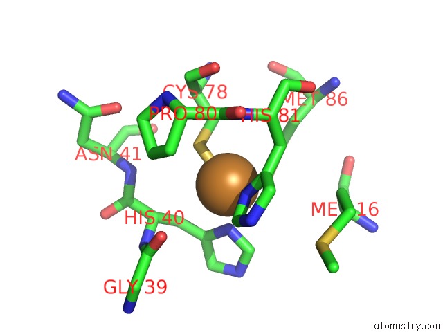
Mono view
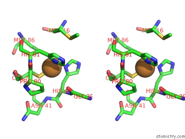
Stereo pair view
Mono view
Stereo pair view
A full contact list of Copper with other atoms in the Cu binding
site number 1 of Refined Crystal Structure of Pseudoazurin From Methylobacterium Extorquens AM1 at 1.5 Angstroms Resolution within 5.0Å range:
|
Reference:
T.Inoue,
Y.Kai,
S.Harada,
N.Kasai,
Y.Ohshiro,
S.Suzuki,
T.Kohzuma,
J.Tobari.
Refined Crystal Structure of Pseudoazurin From Methylobacterium Extorquens AM1 at 1.5 A Resolution. Acta Crystallogr.,Sect.D V. 50 317 1994.
ISSN: ISSN 0907-4449
PubMed: 15299445
DOI: 10.1107/S0907444994000260
Page generated: Mon Jul 14 00:16:57 2025
ISSN: ISSN 0907-4449
PubMed: 15299445
DOI: 10.1107/S0907444994000260
Last articles
I in 2AF6I in 2ANV
I in 2A0N
I in 1XC6
I in 1ZVV
I in 1Z7J
I in 1ZME
I in 1ZCR
I in 1YVP
I in 1YRI