Copper »
PDB 1oe2-1rjp »
1oow »
Copper in PDB 1oow: The Crystal Structure of the Spinach Plastocyanin Double Mutant G8D/L12E Gives Insight Into Its Low Reactivity Towards Photosystem 1 and Cytochrome F
Protein crystallography data
The structure of The Crystal Structure of the Spinach Plastocyanin Double Mutant G8D/L12E Gives Insight Into Its Low Reactivity Towards Photosystem 1 and Cytochrome F, PDB code: 1oow
was solved by
H.Jansson,
M.Okvist,
F.Jacobson,
M.Ejdeback,
O.Hansson,
L.Sjolin,
with X-Ray Crystallography technique. A brief refinement statistics is given in the table below:
| Resolution Low / High (Å) | 19.28 / 2.00 |
| Space group | P 31 2 1 |
| Cell size a, b, c (Å), α, β, γ (°) | 74.761, 74.761, 31.383, 90.00, 90.00, 120.00 |
| R / Rfree (%) | 18.3 / 23.2 |
Copper Binding Sites:
The binding sites of Copper atom in the The Crystal Structure of the Spinach Plastocyanin Double Mutant G8D/L12E Gives Insight Into Its Low Reactivity Towards Photosystem 1 and Cytochrome F
(pdb code 1oow). This binding sites where shown within
5.0 Angstroms radius around Copper atom.
In total only one binding site of Copper was determined in the The Crystal Structure of the Spinach Plastocyanin Double Mutant G8D/L12E Gives Insight Into Its Low Reactivity Towards Photosystem 1 and Cytochrome F, PDB code: 1oow:
In total only one binding site of Copper was determined in the The Crystal Structure of the Spinach Plastocyanin Double Mutant G8D/L12E Gives Insight Into Its Low Reactivity Towards Photosystem 1 and Cytochrome F, PDB code: 1oow:
Copper binding site 1 out of 1 in 1oow
Go back to
Copper binding site 1 out
of 1 in the The Crystal Structure of the Spinach Plastocyanin Double Mutant G8D/L12E Gives Insight Into Its Low Reactivity Towards Photosystem 1 and Cytochrome F
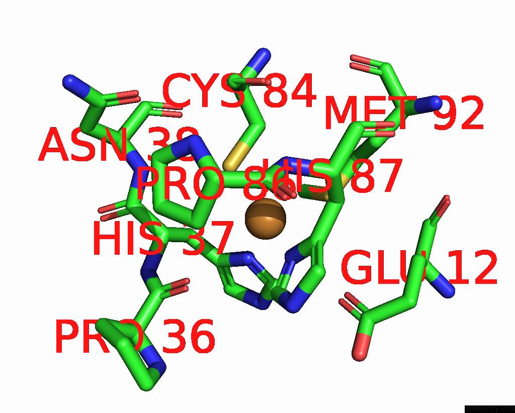
Mono view
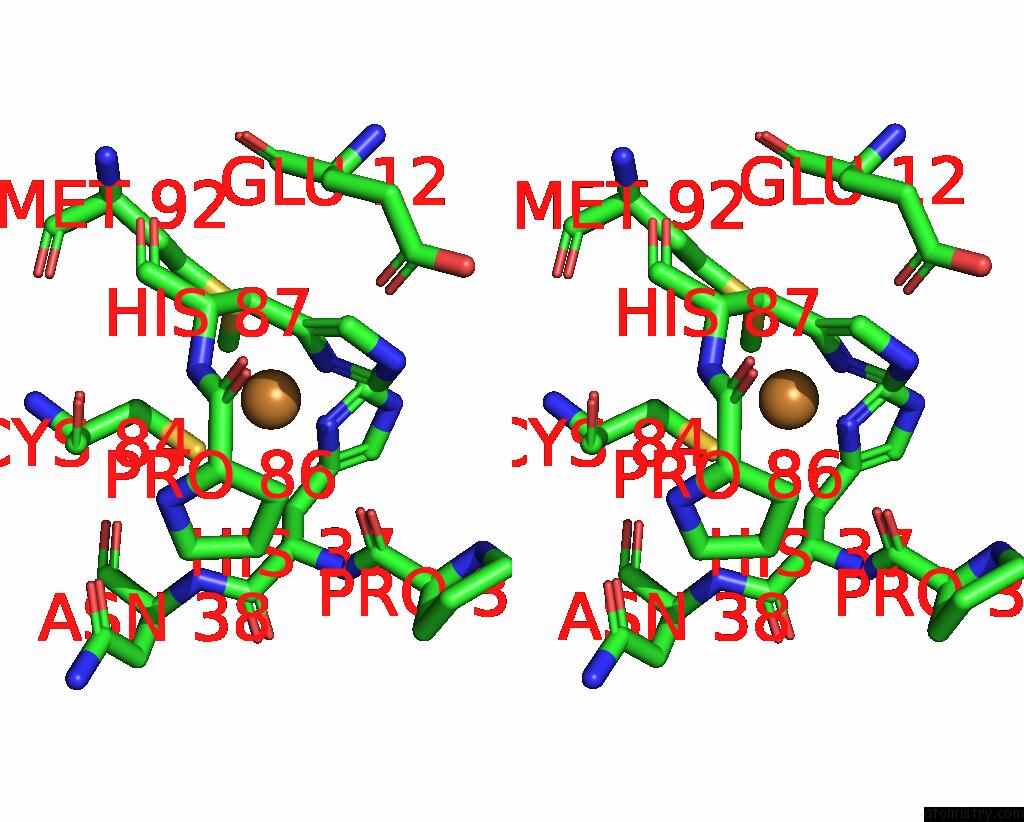
Stereo pair view
Mono view
Stereo pair view
A full contact list of Copper with other atoms in the Cu binding
site number 1 of The Crystal Structure of the Spinach Plastocyanin Double Mutant G8D/L12E Gives Insight Into Its Low Reactivity Towards Photosystem 1 and Cytochrome F within 5.0Å range:
|
Reference:
H.Jansson,
M.Okvist,
F.Jacobson,
M.Ejdeback,
O.Hansson,
L.Sjolin.
The Crystal Structure of the Spinach Plastocyanin Double Mutant G8D/L12E Gives Insight Into Its Low Reactivity Towards Photosystem 1 and Cytochrome F. Biochim.Biophys.Acta V.1607 203 2003.
ISSN: ISSN 0006-3002
PubMed: 14670610
DOI: 10.1016/J.BBABIO.2003.09.011
Page generated: Mon Jul 14 00:15:07 2025
ISSN: ISSN 0006-3002
PubMed: 14670610
DOI: 10.1016/J.BBABIO.2003.09.011
Last articles
I in 3VG2I in 3V6Q
I in 3UVI
I in 3V6N
I in 3V04
I in 3V01
I in 3UVV
I in 3UT8
I in 3UWP
I in 3USM