Copper »
PDB 1a2v-1baw »
1aac »
Copper in PDB 1aac: Amicyanin Oxidized, 1.31 Angstroms
Protein crystallography data
The structure of Amicyanin Oxidized, 1.31 Angstroms, PDB code: 1aac
was solved by
L.M.Cunane,
Z.-W.Chen,
R.C.E.Durley,
F.S.Mathews,
with X-Ray Crystallography technique. A brief refinement statistics is given in the table below:
| Resolution Low / High (Å) | 8.00 / 1.31 |
| Space group | P 1 21 1 |
| Cell size a, b, c (Å), α, β, γ (°) | 28.950, 56.540, 27.550, 90.00, 96.38, 90.00 |
| R / Rfree (%) | 15.5 / n/a |
Copper Binding Sites:
The binding sites of Copper atom in the Amicyanin Oxidized, 1.31 Angstroms
(pdb code 1aac). This binding sites where shown within
5.0 Angstroms radius around Copper atom.
In total only one binding site of Copper was determined in the Amicyanin Oxidized, 1.31 Angstroms, PDB code: 1aac:
In total only one binding site of Copper was determined in the Amicyanin Oxidized, 1.31 Angstroms, PDB code: 1aac:
Copper binding site 1 out of 1 in 1aac
Go back to
Copper binding site 1 out
of 1 in the Amicyanin Oxidized, 1.31 Angstroms
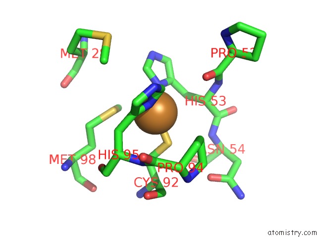
Mono view
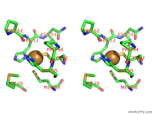
Stereo pair view
Mono view
Stereo pair view
A full contact list of Copper with other atoms in the Cu binding
site number 1 of Amicyanin Oxidized, 1.31 Angstroms within 5.0Å range:
|
Reference:
L.M.Cunane,
Z.W.Chen,
R.C.Durley,
F.S.Mathews.
X-Ray Structure of the Cupredoxin Amicyanin, From Paracoccus Denitrificans, Refined at 1.31 A Resolution. Acta Crystallogr.,Sect.D V. 52 676 1996.
ISSN: ISSN 0907-4449
PubMed: 15299631
DOI: 10.1107/S0907444996001072
Page generated: Sun Jul 13 23:24:32 2025
ISSN: ISSN 0907-4449
PubMed: 15299631
DOI: 10.1107/S0907444996001072
Last articles
K in 6QD4K in 6QM2
K in 6QD3
K in 6QD2
K in 6QD1
K in 6Q6R
K in 6QD0
K in 6QCZ
K in 6QCY
K in 6Q8P