Copper in PDB 8ajz: Serial Femtosecond Crystallography Structure of Co Bound BA3- Type Cytochrome C Oxidase at 2 Milliseconds After Irradiation By A 532 Nm Laser
Enzymatic activity of Serial Femtosecond Crystallography Structure of Co Bound BA3- Type Cytochrome C Oxidase at 2 Milliseconds After Irradiation By A 532 Nm Laser
All present enzymatic activity of Serial Femtosecond Crystallography Structure of Co Bound BA3- Type Cytochrome C Oxidase at 2 Milliseconds After Irradiation By A 532 Nm Laser:
1.9.3.1;
Protein crystallography data
The structure of Serial Femtosecond Crystallography Structure of Co Bound BA3- Type Cytochrome C Oxidase at 2 Milliseconds After Irradiation By A 532 Nm Laser, PDB code: 8ajz
was solved by
C.Safari,
S.Ghosh,
R.Andersson,
J.Johannesson,
A.V.Donoso,
P.Bath,
R.Bosman,
P.Dahl,
E.Nango,
R.Tanaka,
D.Zoric,
E.Svensson,
T.Nakane,
S.Iwata,
R.Neutze,
G.Branden,
with X-Ray Crystallography technique. A brief refinement statistics is given in the table below:
Resolution Low / High (Å)
|
38.70 /
2.00
|
Space group
|
C 1 2 1
|
Cell size a, b, c (Å), α, β, γ (°)
|
145.85,
100.32,
96.62,
90,
126.76,
90
|
R / Rfree (%)
|
19 /
21
|
Other elements in 8ajz:
The structure of Serial Femtosecond Crystallography Structure of Co Bound BA3- Type Cytochrome C Oxidase at 2 Milliseconds After Irradiation By A 532 Nm Laser also contains other interesting chemical elements:
Copper Binding Sites:
The binding sites of Copper atom in the Serial Femtosecond Crystallography Structure of Co Bound BA3- Type Cytochrome C Oxidase at 2 Milliseconds After Irradiation By A 532 Nm Laser
(pdb code 8ajz). This binding sites where shown within
5.0 Angstroms radius around Copper atom.
In total 4 binding sites of Copper where determined in the
Serial Femtosecond Crystallography Structure of Co Bound BA3- Type Cytochrome C Oxidase at 2 Milliseconds After Irradiation By A 532 Nm Laser, PDB code: 8ajz:
Jump to Copper binding site number:
1;
2;
3;
4;
Copper binding site 1 out
of 4 in 8ajz
Go back to
Copper Binding Sites List in 8ajz
Copper binding site 1 out
of 4 in the Serial Femtosecond Crystallography Structure of Co Bound BA3- Type Cytochrome C Oxidase at 2 Milliseconds After Irradiation By A 532 Nm Laser
Mono view
Stereo pair view
|
A full contact list of Copper with other atoms in the Cu binding
site number 1 of Serial Femtosecond Crystallography Structure of Co Bound BA3- Type Cytochrome C Oxidase at 2 Milliseconds After Irradiation By A 532 Nm Laser within 5.0Å range:
probe
|
atom
|
residue
|
distance (Å)
|
B
|
Occ
|
A:Cu601
b:30.6
occ:0.70
|
CU
|
A:CU601
|
0.0
|
30.6
|
0.7
|
CU
|
A:CU601
|
0.3
|
46.4
|
0.3
|
NE2
|
A:HIS283
|
1.9
|
46.4
|
0.3
|
NE2
|
A:HIS282
|
2.0
|
43.5
|
0.3
|
ND1
|
A:HIS233
|
2.0
|
36.7
|
0.3
|
NE2
|
A:HIS282
|
2.1
|
29.0
|
0.7
|
ND1
|
A:HIS233
|
2.1
|
27.1
|
0.7
|
NE2
|
A:HIS283
|
2.1
|
27.6
|
0.7
|
C
|
A:CMO614
|
2.3
|
43.5
|
0.3
|
O
|
A:CMO614
|
2.3
|
28.4
|
0.7
|
O
|
A:CMO614
|
2.5
|
35.6
|
0.3
|
CE1
|
A:HIS283
|
2.5
|
44.1
|
0.3
|
CE1
|
A:HIS282
|
2.8
|
42.3
|
0.3
|
CG
|
A:HIS233
|
2.8
|
40.1
|
0.3
|
CG
|
A:HIS233
|
3.0
|
26.1
|
0.7
|
CE1
|
A:HIS283
|
3.0
|
27.1
|
0.7
|
CE1
|
A:HIS282
|
3.0
|
28.2
|
0.7
|
CB
|
A:HIS233
|
3.0
|
39.6
|
0.3
|
CE1
|
A:HIS233
|
3.1
|
26.2
|
0.7
|
CD2
|
A:HIS282
|
3.1
|
27.1
|
0.7
|
CD2
|
A:HIS283
|
3.1
|
44.6
|
0.3
|
CD2
|
A:HIS283
|
3.1
|
27.7
|
0.7
|
CD2
|
A:HIS282
|
3.1
|
45.1
|
0.3
|
CE1
|
A:HIS233
|
3.1
|
37.6
|
0.3
|
C
|
A:CMO614
|
3.2
|
26.4
|
0.7
|
CB
|
A:HIS233
|
3.3
|
25.8
|
0.7
|
CA
|
A:HIS233
|
3.6
|
40.6
|
0.3
|
CA
|
A:HIS233
|
3.7
|
25.9
|
0.7
|
ND1
|
A:HIS283
|
3.7
|
44.4
|
0.3
|
ND1
|
A:HIS282
|
4.0
|
45.4
|
0.3
|
CD2
|
A:HIS233
|
4.0
|
41.9
|
0.3
|
CG
|
A:HIS283
|
4.0
|
46.7
|
0.3
|
NE2
|
A:HIS233
|
4.1
|
41.0
|
0.3
|
ND1
|
A:HIS283
|
4.1
|
26.4
|
0.7
|
ND1
|
A:HIS282
|
4.2
|
27.8
|
0.7
|
CG
|
A:HIS282
|
4.2
|
46.1
|
0.3
|
O
|
A:HOH713
|
4.2
|
25.7
|
0.3
|
CD2
|
A:HIS233
|
4.2
|
26.6
|
0.7
|
CG
|
A:HIS283
|
4.2
|
26.8
|
0.7
|
NE2
|
A:HIS233
|
4.2
|
27.8
|
0.7
|
CG
|
A:HIS282
|
4.2
|
26.8
|
0.7
|
C1D
|
A:HAS603
|
4.3
|
53.9
|
0.3
|
ND
|
A:HAS603
|
4.3
|
32.2
|
0.7
|
C1D
|
A:HAS603
|
4.3
|
31.3
|
0.7
|
C2D
|
A:HAS603
|
4.4
|
51.1
|
0.3
|
ND
|
A:HAS603
|
4.4
|
48.0
|
0.3
|
C4D
|
A:HAS603
|
4.4
|
31.2
|
0.7
|
C4D
|
A:HAS603
|
4.5
|
44.9
|
0.3
|
C2D
|
A:HAS603
|
4.5
|
30.3
|
0.7
|
C3D
|
A:HAS603
|
4.6
|
45.4
|
0.3
|
N
|
A:HIS233
|
4.6
|
34.6
|
1.0
|
C3D
|
A:HAS603
|
4.7
|
29.9
|
0.7
|
C
|
A:HIS233
|
4.7
|
32.1
|
1.0
|
OMD
|
A:HAS603
|
4.7
|
62.4
|
0.3
|
CHB
|
A:HAS603
|
4.7
|
31.1
|
0.7
|
O
|
A:HIS233
|
4.8
|
32.4
|
1.0
|
CHB
|
A:HAS603
|
4.8
|
47.9
|
0.3
|
O
|
A:GLY232
|
4.9
|
29.8
|
1.0
|
FE
|
A:HAS603
|
4.9
|
35.2
|
0.7
|
CHA
|
A:HAS603
|
5.0
|
30.1
|
0.7
|
|
Copper binding site 2 out
of 4 in 8ajz
Go back to
Copper Binding Sites List in 8ajz
Copper binding site 2 out
of 4 in the Serial Femtosecond Crystallography Structure of Co Bound BA3- Type Cytochrome C Oxidase at 2 Milliseconds After Irradiation By A 532 Nm Laser
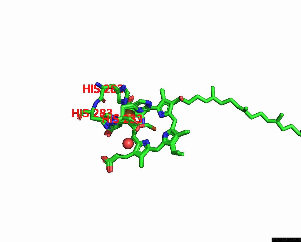
Mono view
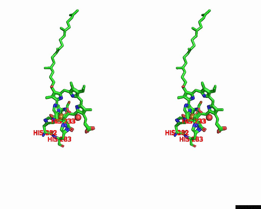
Stereo pair view
|
A full contact list of Copper with other atoms in the Cu binding
site number 2 of Serial Femtosecond Crystallography Structure of Co Bound BA3- Type Cytochrome C Oxidase at 2 Milliseconds After Irradiation By A 532 Nm Laser within 5.0Å range:
probe
|
atom
|
residue
|
distance (Å)
|
B
|
Occ
|
A:Cu601
b:46.4
occ:0.30
|
CU
|
A:CU601
|
0.0
|
46.4
|
0.3
|
CU
|
A:CU601
|
0.3
|
30.6
|
0.7
|
NE2
|
A:HIS282
|
2.0
|
43.5
|
0.3
|
C
|
A:CMO614
|
2.0
|
43.5
|
0.3
|
NE2
|
A:HIS283
|
2.0
|
46.4
|
0.3
|
ND1
|
A:HIS233
|
2.1
|
36.7
|
0.3
|
NE2
|
A:HIS282
|
2.1
|
29.0
|
0.7
|
O
|
A:CMO614
|
2.1
|
28.4
|
0.7
|
ND1
|
A:HIS233
|
2.2
|
27.1
|
0.7
|
NE2
|
A:HIS283
|
2.2
|
27.6
|
0.7
|
O
|
A:CMO614
|
2.3
|
35.6
|
0.3
|
CE1
|
A:HIS283
|
2.5
|
44.1
|
0.3
|
CE1
|
A:HIS282
|
2.8
|
42.3
|
0.3
|
CG
|
A:HIS233
|
2.9
|
40.1
|
0.3
|
C
|
A:CMO614
|
2.9
|
26.4
|
0.7
|
CE1
|
A:HIS283
|
3.0
|
27.1
|
0.7
|
CE1
|
A:HIS233
|
3.0
|
26.2
|
0.7
|
CE1
|
A:HIS233
|
3.1
|
37.6
|
0.3
|
CE1
|
A:HIS282
|
3.1
|
28.2
|
0.7
|
CD2
|
A:HIS282
|
3.1
|
27.1
|
0.7
|
CD2
|
A:HIS282
|
3.1
|
45.1
|
0.3
|
CG
|
A:HIS233
|
3.2
|
26.1
|
0.7
|
CB
|
A:HIS233
|
3.3
|
39.6
|
0.3
|
CD2
|
A:HIS283
|
3.3
|
27.7
|
0.7
|
CD2
|
A:HIS283
|
3.3
|
44.6
|
0.3
|
CB
|
A:HIS233
|
3.5
|
25.8
|
0.7
|
ND1
|
A:HIS283
|
3.8
|
44.4
|
0.3
|
CA
|
A:HIS233
|
3.9
|
40.6
|
0.3
|
CA
|
A:HIS233
|
3.9
|
25.9
|
0.7
|
ND
|
A:HAS603
|
4.0
|
32.2
|
0.7
|
ND1
|
A:HIS282
|
4.0
|
45.4
|
0.3
|
C1D
|
A:HAS603
|
4.0
|
53.9
|
0.3
|
C1D
|
A:HAS603
|
4.0
|
31.3
|
0.7
|
CD2
|
A:HIS233
|
4.1
|
41.9
|
0.3
|
NE2
|
A:HIS233
|
4.1
|
41.0
|
0.3
|
O
|
A:HOH713
|
4.2
|
25.7
|
0.3
|
CG
|
A:HIS283
|
4.2
|
46.7
|
0.3
|
ND1
|
A:HIS283
|
4.2
|
26.4
|
0.7
|
ND
|
A:HAS603
|
4.2
|
48.0
|
0.3
|
CG
|
A:HIS282
|
4.2
|
46.1
|
0.3
|
ND1
|
A:HIS282
|
4.2
|
27.8
|
0.7
|
NE2
|
A:HIS233
|
4.2
|
27.8
|
0.7
|
C4D
|
A:HAS603
|
4.2
|
31.2
|
0.7
|
C2D
|
A:HAS603
|
4.2
|
51.1
|
0.3
|
CG
|
A:HIS282
|
4.2
|
26.8
|
0.7
|
CD2
|
A:HIS233
|
4.3
|
26.6
|
0.7
|
C4D
|
A:HAS603
|
4.3
|
44.9
|
0.3
|
CG
|
A:HIS283
|
4.3
|
26.8
|
0.7
|
C2D
|
A:HAS603
|
4.4
|
30.3
|
0.7
|
C3D
|
A:HAS603
|
4.4
|
45.4
|
0.3
|
C3D
|
A:HAS603
|
4.5
|
29.9
|
0.7
|
CHB
|
A:HAS603
|
4.5
|
31.1
|
0.7
|
CHB
|
A:HAS603
|
4.6
|
47.9
|
0.3
|
OMD
|
A:HAS603
|
4.6
|
62.4
|
0.3
|
FE
|
A:HAS603
|
4.6
|
35.2
|
0.7
|
CHA
|
A:HAS603
|
4.8
|
30.1
|
0.7
|
N
|
A:HIS233
|
4.8
|
34.6
|
1.0
|
C
|
A:HIS233
|
4.9
|
32.1
|
1.0
|
CMD
|
A:HAS603
|
4.9
|
62.1
|
0.3
|
O
|
A:HIS233
|
4.9
|
32.4
|
1.0
|
FE
|
A:HAS603
|
4.9
|
49.9
|
0.3
|
C1B
|
A:HAS603
|
4.9
|
31.9
|
0.7
|
NB
|
A:HAS603
|
4.9
|
32.8
|
0.7
|
NA
|
A:HAS603
|
5.0
|
31.2
|
0.7
|
C1B
|
A:HAS603
|
5.0
|
51.5
|
0.3
|
|
Copper binding site 3 out
of 4 in 8ajz
Go back to
Copper Binding Sites List in 8ajz
Copper binding site 3 out
of 4 in the Serial Femtosecond Crystallography Structure of Co Bound BA3- Type Cytochrome C Oxidase at 2 Milliseconds After Irradiation By A 532 Nm Laser
Mono view
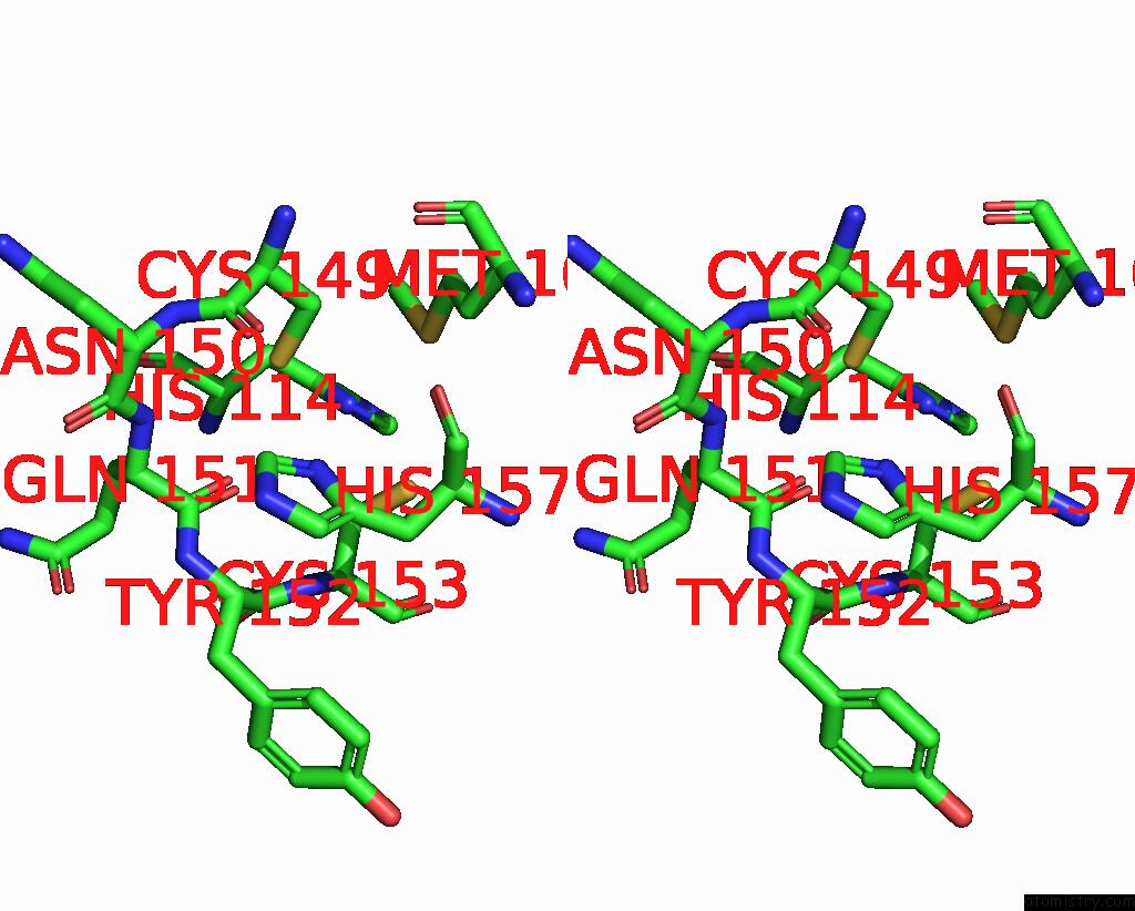
Stereo pair view
|
A full contact list of Copper with other atoms in the Cu binding
site number 3 of Serial Femtosecond Crystallography Structure of Co Bound BA3- Type Cytochrome C Oxidase at 2 Milliseconds After Irradiation By A 532 Nm Laser within 5.0Å range:
probe
|
atom
|
residue
|
distance (Å)
|
B
|
Occ
|
B:Cu203
b:33.3
occ:1.00
|
CU1
|
B:CUA203
|
0.0
|
33.3
|
1.0
|
ND1
|
B:HIS157
|
2.0
|
26.6
|
1.0
|
O
|
B:GLN151
|
2.5
|
26.1
|
1.0
|
SG
|
B:CYS149
|
2.5
|
29.5
|
1.0
|
CU2
|
B:CUA203
|
2.6
|
34.6
|
1.0
|
CE1
|
B:HIS157
|
2.8
|
27.9
|
1.0
|
SG
|
B:CYS153
|
2.9
|
36.5
|
1.0
|
CG
|
B:HIS157
|
3.1
|
25.6
|
1.0
|
CB
|
B:CYS153
|
3.3
|
37.8
|
1.0
|
CB
|
B:CYS149
|
3.4
|
31.9
|
1.0
|
C
|
B:GLN151
|
3.5
|
27.7
|
1.0
|
CB
|
B:HIS157
|
3.5
|
26.4
|
1.0
|
N
|
B:CYS153
|
3.6
|
37.9
|
1.0
|
CA
|
B:HIS157
|
3.8
|
28.1
|
1.0
|
NE2
|
B:HIS157
|
4.0
|
27.6
|
1.0
|
CA
|
B:CYS153
|
4.1
|
37.6
|
1.0
|
O
|
B:CYS149
|
4.1
|
29.9
|
1.0
|
CD2
|
B:HIS157
|
4.1
|
26.9
|
1.0
|
C
|
B:TYR152
|
4.2
|
30.6
|
1.0
|
O
|
B:HIS157
|
4.2
|
28.7
|
1.0
|
CA
|
B:TYR152
|
4.2
|
30.3
|
1.0
|
N
|
B:TYR152
|
4.2
|
28.8
|
1.0
|
C
|
B:CYS149
|
4.3
|
30.9
|
1.0
|
N
|
B:GLN151
|
4.3
|
29.4
|
1.0
|
ND1
|
B:HIS114
|
4.4
|
25.7
|
1.0
|
C
|
B:HIS157
|
4.4
|
29.5
|
1.0
|
SD
|
B:MET160
|
4.5
|
29.7
|
1.0
|
CA
|
B:CYS149
|
4.5
|
30.5
|
1.0
|
CA
|
B:GLN151
|
4.5
|
29.5
|
1.0
|
CB
|
B:MET160
|
4.8
|
30.0
|
1.0
|
N
|
B:ASN150
|
4.9
|
28.5
|
1.0
|
N
|
B:HIS157
|
5.0
|
29.9
|
1.0
|
|
Copper binding site 4 out
of 4 in 8ajz
Go back to
Copper Binding Sites List in 8ajz
Copper binding site 4 out
of 4 in the Serial Femtosecond Crystallography Structure of Co Bound BA3- Type Cytochrome C Oxidase at 2 Milliseconds After Irradiation By A 532 Nm Laser
Mono view
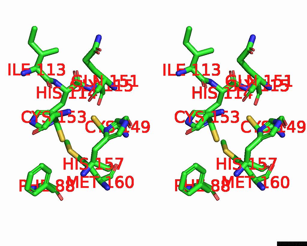
Stereo pair view
|
A full contact list of Copper with other atoms in the Cu binding
site number 4 of Serial Femtosecond Crystallography Structure of Co Bound BA3- Type Cytochrome C Oxidase at 2 Milliseconds After Irradiation By A 532 Nm Laser within 5.0Å range:
probe
|
atom
|
residue
|
distance (Å)
|
B
|
Occ
|
B:Cu203
b:34.6
occ:1.00
|
CU2
|
B:CUA203
|
0.0
|
34.6
|
1.0
|
ND1
|
B:HIS114
|
2.1
|
25.7
|
1.0
|
SG
|
B:CYS149
|
2.4
|
29.5
|
1.0
|
SD
|
B:MET160
|
2.4
|
29.7
|
1.0
|
CU1
|
B:CUA203
|
2.6
|
33.3
|
1.0
|
SG
|
B:CYS153
|
2.7
|
36.5
|
1.0
|
CE1
|
B:HIS114
|
2.9
|
27.1
|
1.0
|
CB
|
B:CYS153
|
3.1
|
37.8
|
1.0
|
CG
|
B:HIS114
|
3.2
|
28.3
|
1.0
|
CE
|
B:MET160
|
3.2
|
28.8
|
1.0
|
CB
|
B:CYS149
|
3.3
|
31.9
|
1.0
|
CB
|
B:HIS114
|
3.6
|
28.6
|
1.0
|
CG
|
B:MET160
|
3.8
|
32.6
|
1.0
|
CA
|
B:HIS114
|
4.0
|
30.7
|
1.0
|
O
|
B:GLN151
|
4.0
|
26.1
|
1.0
|
NE2
|
B:HIS114
|
4.1
|
26.0
|
1.0
|
CB
|
B:MET160
|
4.2
|
30.0
|
1.0
|
CD2
|
B:HIS114
|
4.2
|
27.4
|
1.0
|
ND1
|
B:HIS157
|
4.5
|
26.6
|
1.0
|
CA
|
B:CYS153
|
4.6
|
37.6
|
1.0
|
CA
|
B:CYS149
|
4.7
|
30.5
|
1.0
|
O
|
B:ILE113
|
4.7
|
28.9
|
1.0
|
N
|
B:GLY115
|
4.8
|
30.8
|
1.0
|
CD1
|
B:PHE88
|
4.9
|
31.0
|
1.0
|
N
|
B:CYS153
|
4.9
|
37.9
|
1.0
|
|
Reference:
C.Safari,
S.Ghosh,
R.Andersson,
J.Johannesson,
A.V.Donoso,
P.Bath,
R.Bosman,
P.Dahl,
E.Nango,
R.Tanaka,
D.Zoric,
E.Svensson,
T.Nakane,
S.Iwata,
R.Neutze,
G.Branden.
Serial Femtosecond Crystallography Structure of Co Bound BA3- Type Cytochrome C Oxidase at 2 Milliseconds After Irradiation By A 532 Nm Laser To Be Published.
Page generated: Wed Jul 31 09:31:32 2024
|