Copper »
PDB 7xmc-8b9r »
7zjb »
Copper in PDB 7zjb: Structural and Functional Characterization of the Bacterial Lytic Polysaccharide Monooxygenase SCLPMO10D
Protein crystallography data
The structure of Structural and Functional Characterization of the Bacterial Lytic Polysaccharide Monooxygenase SCLPMO10D, PDB code: 7zjb
was solved by
A.K.Votvik,
A.K.Rohr,
A.A.Stepnov,
B.Bissaro,
M.Sorlie,
V.G.H.Eijsink,
Z.Forsberg,
with X-Ray Crystallography technique. A brief refinement statistics is given in the table below:
| Resolution Low / High (Å) | 54.81 / 1.37 |
| Space group | P 43 21 2 |
| Cell size a, b, c (Å), α, β, γ (°) | 59.08, 59.08, 145.718, 90, 90, 90 |
| R / Rfree (%) | 12.9 / 15.2 |
Other elements in 7zjb:
The structure of Structural and Functional Characterization of the Bacterial Lytic Polysaccharide Monooxygenase SCLPMO10D also contains other interesting chemical elements:
| Sodium | (Na) | 1 atom |
Copper Binding Sites:
The binding sites of Copper atom in the Structural and Functional Characterization of the Bacterial Lytic Polysaccharide Monooxygenase SCLPMO10D
(pdb code 7zjb). This binding sites where shown within
5.0 Angstroms radius around Copper atom.
In total only one binding site of Copper was determined in the Structural and Functional Characterization of the Bacterial Lytic Polysaccharide Monooxygenase SCLPMO10D, PDB code: 7zjb:
In total only one binding site of Copper was determined in the Structural and Functional Characterization of the Bacterial Lytic Polysaccharide Monooxygenase SCLPMO10D, PDB code: 7zjb:
Copper binding site 1 out of 1 in 7zjb
Go back to
Copper binding site 1 out
of 1 in the Structural and Functional Characterization of the Bacterial Lytic Polysaccharide Monooxygenase SCLPMO10D

Mono view
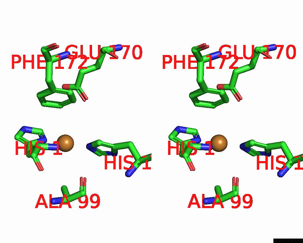
Stereo pair view
Mono view
Stereo pair view
A full contact list of Copper with other atoms in the Cu binding
site number 1 of Structural and Functional Characterization of the Bacterial Lytic Polysaccharide Monooxygenase SCLPMO10D within 5.0Å range:
|
Reference:
A.K.Votvik,
A.K.Rohr,
B.Bissaro,
A.A.Stepnov,
M.Sorlie,
V.G.H.Eijsink,
Z.Forsberg.
Structural and Functional Characterization of the Catalytic Domain of A Cell-Wall Anchored Bacterial Lytic Polysaccharide Monooxygenase From Streptomyces Coelicolor. Sci Rep V. 13 5345 2023.
ISSN: ESSN 2045-2322
PubMed: 37005446
DOI: 10.1038/S41598-023-32263-7
Page generated: Mon Jul 14 08:46:21 2025
ISSN: ESSN 2045-2322
PubMed: 37005446
DOI: 10.1038/S41598-023-32263-7
Last articles
Fe in 2YXOFe in 2YRS
Fe in 2YXC
Fe in 2YNM
Fe in 2YVJ
Fe in 2YP1
Fe in 2YU2
Fe in 2YU1
Fe in 2YQB
Fe in 2YOO