Copper »
PDB 7s1f-7xmb »
7xma »
Copper in PDB 7xma: Crystal Structure of Bovine Heart Cytochrome C Oxidase, Apo Structure with Dmso
Enzymatic activity of Crystal Structure of Bovine Heart Cytochrome C Oxidase, Apo Structure with Dmso
All present enzymatic activity of Crystal Structure of Bovine Heart Cytochrome C Oxidase, Apo Structure with Dmso:
7.1.1.9;
7.1.1.9;
Protein crystallography data
The structure of Crystal Structure of Bovine Heart Cytochrome C Oxidase, Apo Structure with Dmso, PDB code: 7xma
was solved by
Y.Nishida,
K.Shinzawa-Itoh,
N.Mizuno,
T.Kumasaka,
S.Yoshikawa,
T.Tsukihara,
S.Takashima,
Y.Shintani,
with X-Ray Crystallography technique. A brief refinement statistics is given in the table below:
| Resolution Low / High (Å) | 29.97 / 2.20 |
| Space group | P 21 21 21 |
| Cell size a, b, c (Å), α, β, γ (°) | 181.8, 203.577, 177.859, 90, 90, 90 |
| R / Rfree (%) | 18.6 / 21.3 |
Other elements in 7xma:
The structure of Crystal Structure of Bovine Heart Cytochrome C Oxidase, Apo Structure with Dmso also contains other interesting chemical elements:
| Iron | (Fe) | 4 atoms |
| Zinc | (Zn) | 2 atoms |
| Sodium | (Na) | 2 atoms |
| Magnesium | (Mg) | 2 atoms |
Copper Binding Sites:
The binding sites of Copper atom in the Crystal Structure of Bovine Heart Cytochrome C Oxidase, Apo Structure with Dmso
(pdb code 7xma). This binding sites where shown within
5.0 Angstroms radius around Copper atom.
In total 6 binding sites of Copper where determined in the Crystal Structure of Bovine Heart Cytochrome C Oxidase, Apo Structure with Dmso, PDB code: 7xma:
Jump to Copper binding site number: 1; 2; 3; 4; 5; 6;
In total 6 binding sites of Copper where determined in the Crystal Structure of Bovine Heart Cytochrome C Oxidase, Apo Structure with Dmso, PDB code: 7xma:
Jump to Copper binding site number: 1; 2; 3; 4; 5; 6;
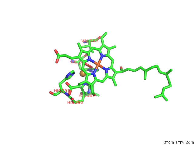
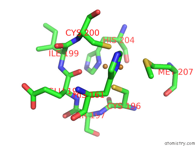
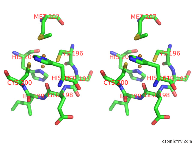
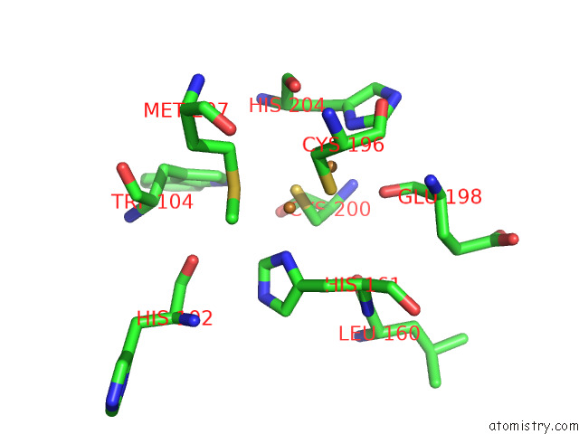
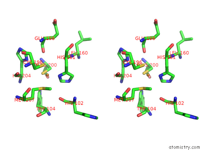
Copper binding site 1 out of 6 in 7xma
Go back to
Copper binding site 1 out
of 6 in the Crystal Structure of Bovine Heart Cytochrome C Oxidase, Apo Structure with Dmso
Mono view
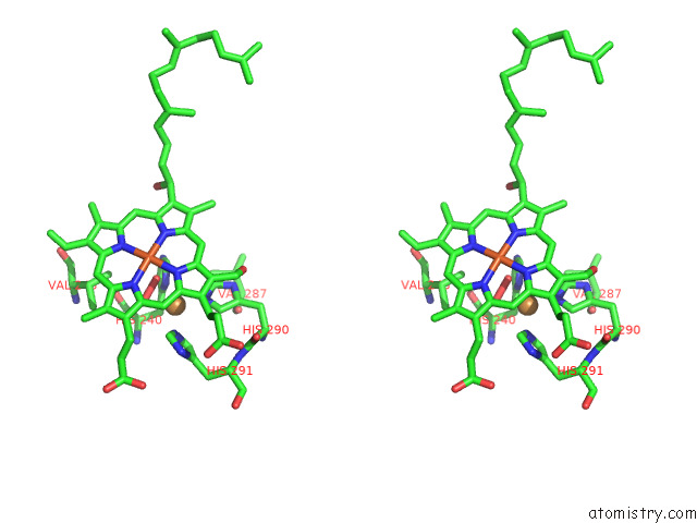
Stereo pair view
Mono view
Stereo pair view
A full contact list of Copper with other atoms in the Cu binding
site number 1 of Crystal Structure of Bovine Heart Cytochrome C Oxidase, Apo Structure with Dmso within 5.0Å range:
|
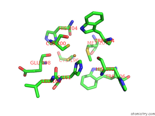
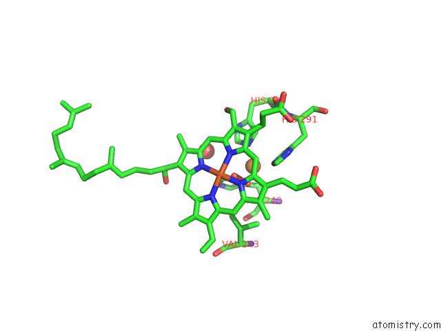
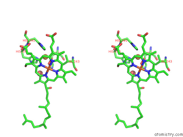
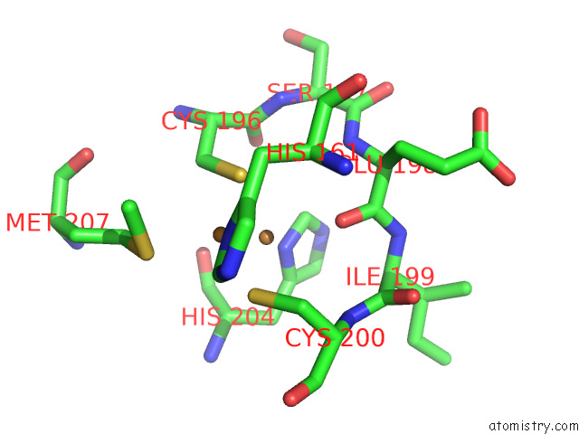
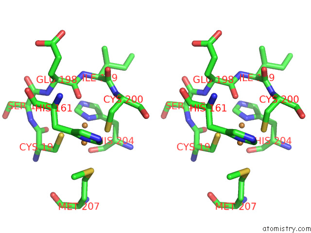
Copper binding site 2 out of 6 in 7xma
Go back to
Copper binding site 2 out
of 6 in the Crystal Structure of Bovine Heart Cytochrome C Oxidase, Apo Structure with Dmso
Mono view
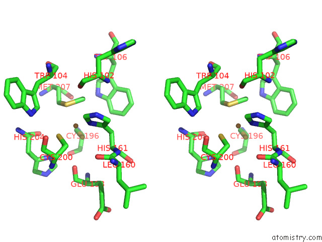
Stereo pair view
Mono view
Stereo pair view
A full contact list of Copper with other atoms in the Cu binding
site number 2 of Crystal Structure of Bovine Heart Cytochrome C Oxidase, Apo Structure with Dmso within 5.0Å range:
|
Copper binding site 3 out of 6 in 7xma
Go back to
Copper binding site 3 out
of 6 in the Crystal Structure of Bovine Heart Cytochrome C Oxidase, Apo Structure with Dmso
Mono view
Stereo pair view
Mono view
Stereo pair view
A full contact list of Copper with other atoms in the Cu binding
site number 3 of Crystal Structure of Bovine Heart Cytochrome C Oxidase, Apo Structure with Dmso within 5.0Å range:
|
Copper binding site 4 out of 6 in 7xma
Go back to
Copper binding site 4 out
of 6 in the Crystal Structure of Bovine Heart Cytochrome C Oxidase, Apo Structure with Dmso
Mono view
Stereo pair view
Mono view
Stereo pair view
A full contact list of Copper with other atoms in the Cu binding
site number 4 of Crystal Structure of Bovine Heart Cytochrome C Oxidase, Apo Structure with Dmso within 5.0Å range:
|
Copper binding site 5 out of 6 in 7xma
Go back to
Copper binding site 5 out
of 6 in the Crystal Structure of Bovine Heart Cytochrome C Oxidase, Apo Structure with Dmso
Mono view
Stereo pair view
Mono view
Stereo pair view
A full contact list of Copper with other atoms in the Cu binding
site number 5 of Crystal Structure of Bovine Heart Cytochrome C Oxidase, Apo Structure with Dmso within 5.0Å range:
|
Copper binding site 6 out of 6 in 7xma
Go back to
Copper binding site 6 out
of 6 in the Crystal Structure of Bovine Heart Cytochrome C Oxidase, Apo Structure with Dmso
Mono view
Stereo pair view
Mono view
Stereo pair view
A full contact list of Copper with other atoms in the Cu binding
site number 6 of Crystal Structure of Bovine Heart Cytochrome C Oxidase, Apo Structure with Dmso within 5.0Å range:
|
Reference:
Y.Nishida,
S.Yanagisawa,
R.Morita,
H.Shigematsu,
K.Shinzawa-Itoh,
H.Yuki,
S.Ogasawara,
K.Shimuta,
T.Iwamoto,
C.Nakabayashi,
W.Matsumura,
H.Kato,
C.Gopalasingam,
T.Nagao,
T.Qaqorh,
Y.Takahashi,
S.Yamazaki,
K.Kamiya,
R.Harada,
N.Mizuno,
H.Takahashi,
Y.Akeda,
M.Ohnishi,
Y.Ishii,
T.Kumasaka,
T.Murata,
K.Muramoto,
T.Tosha,
Y.Shiro,
T.Honma,
Y.Shigeta,
M.Kubo,
S.Takashima,
Y.Shintani.
Identifying Antibiotics Based on Structural Differences in the Conserved Allostery From Mitochondrial Heme-Copper Oxidases. Nat Commun V. 13 7591 2022.
ISSN: ESSN 2041-1723
PubMed: 36481732
DOI: 10.1038/S41467-022-34771-Y
Page generated: Wed Jul 31 09:22:31 2024
ISSN: ESSN 2041-1723
PubMed: 36481732
DOI: 10.1038/S41467-022-34771-Y
Last articles
Ca in 5SZLCa in 5SY1
Ca in 5SWI
Ca in 5SVE
Ca in 5SSX
Ca in 5SV0
Ca in 5STD
Ca in 5SSZ
Ca in 5SSY
Ca in 5SIC