Copper »
PDB 7s1f-7xmb »
7vw1 »
Copper in PDB 7vw1: Structure of A Dimeric Periplasmic Protein Bound with Cuprous Ions
Protein crystallography data
The structure of Structure of A Dimeric Periplasmic Protein Bound with Cuprous Ions, PDB code: 7vw1
was solved by
J.Yang,
L.Liu,
with X-Ray Crystallography technique. A brief refinement statistics is given in the table below:
| Resolution Low / High (Å) | 34.62 / 2.49 |
| Space group | P 43 |
| Cell size a, b, c (Å), α, β, γ (°) | 77.415, 77.415, 44.933, 90, 90, 90 |
| R / Rfree (%) | 20.3 / 24.8 |
Copper Binding Sites:
The binding sites of Copper atom in the Structure of A Dimeric Periplasmic Protein Bound with Cuprous Ions
(pdb code 7vw1). This binding sites where shown within
5.0 Angstroms radius around Copper atom.
In total 5 binding sites of Copper where determined in the Structure of A Dimeric Periplasmic Protein Bound with Cuprous Ions, PDB code: 7vw1:
Jump to Copper binding site number: 1; 2; 3; 4; 5;
In total 5 binding sites of Copper where determined in the Structure of A Dimeric Periplasmic Protein Bound with Cuprous Ions, PDB code: 7vw1:
Jump to Copper binding site number: 1; 2; 3; 4; 5;
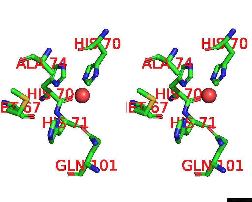
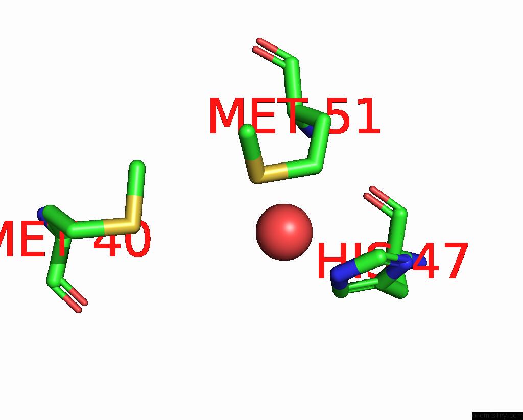
Copper binding site 1 out of 5 in 7vw1
Go back to
Copper binding site 1 out
of 5 in the Structure of A Dimeric Periplasmic Protein Bound with Cuprous Ions

Mono view
Stereo pair view
Mono view
Stereo pair view
A full contact list of Copper with other atoms in the Cu binding
site number 1 of Structure of A Dimeric Periplasmic Protein Bound with Cuprous Ions within 5.0Å range:
|
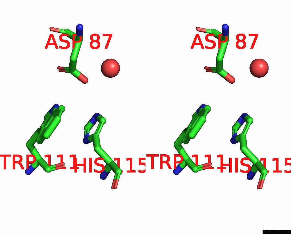
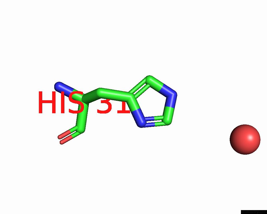
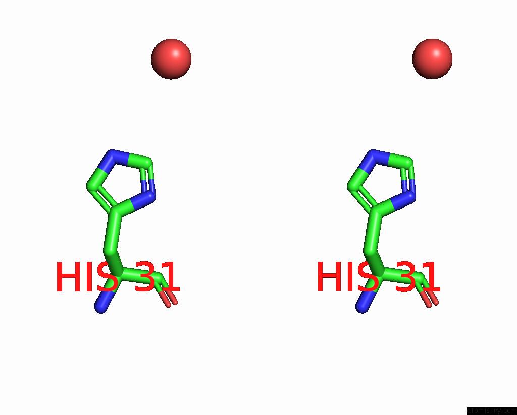
Copper binding site 2 out of 5 in 7vw1
Go back to
Copper binding site 2 out
of 5 in the Structure of A Dimeric Periplasmic Protein Bound with Cuprous Ions

Mono view
Stereo pair view
Mono view
Stereo pair view
A full contact list of Copper with other atoms in the Cu binding
site number 2 of Structure of A Dimeric Periplasmic Protein Bound with Cuprous Ions within 5.0Å range:
|
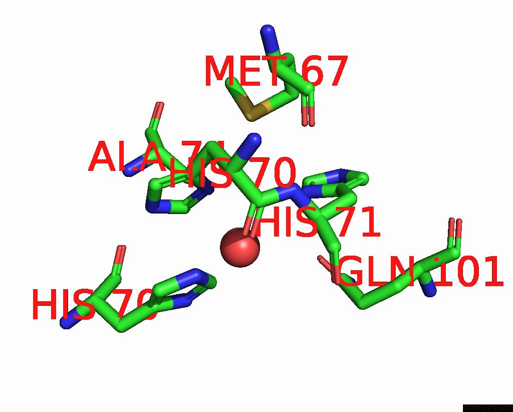
Copper binding site 3 out of 5 in 7vw1
Go back to
Copper binding site 3 out
of 5 in the Structure of A Dimeric Periplasmic Protein Bound with Cuprous Ions
Mono view
Stereo pair view
Mono view
Stereo pair view
A full contact list of Copper with other atoms in the Cu binding
site number 3 of Structure of A Dimeric Periplasmic Protein Bound with Cuprous Ions within 5.0Å range:
|
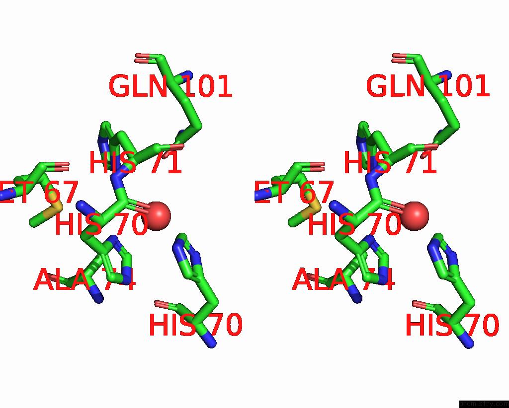
Copper binding site 4 out of 5 in 7vw1
Go back to
Copper binding site 4 out
of 5 in the Structure of A Dimeric Periplasmic Protein Bound with Cuprous Ions
Mono view

Stereo pair view
Mono view
Stereo pair view
A full contact list of Copper with other atoms in the Cu binding
site number 4 of Structure of A Dimeric Periplasmic Protein Bound with Cuprous Ions within 5.0Å range:
|
Copper binding site 5 out of 5 in 7vw1
Go back to
Copper binding site 5 out
of 5 in the Structure of A Dimeric Periplasmic Protein Bound with Cuprous Ions
Mono view
Stereo pair view
Mono view
Stereo pair view
A full contact list of Copper with other atoms in the Cu binding
site number 5 of Structure of A Dimeric Periplasmic Protein Bound with Cuprous Ions within 5.0Å range:
|
Reference:
J.Yang,
M.Gao,
J.Wang,
C.He,
X.Wang,
L.Liu.
Structural Basis of Copper Binding By A Dimeric Periplasmic Protein Forming A Six-Helical Bundle. J.Inorg.Biochem. V. 229 11728 2022.
ISSN: ISSN 0162-0134
PubMed: 35066349
DOI: 10.1016/J.JINORGBIO.2022.111728
Page generated: Wed Jul 31 09:18:25 2024
ISSN: ISSN 0162-0134
PubMed: 35066349
DOI: 10.1016/J.JINORGBIO.2022.111728
Last articles
Zn in 9JYWZn in 9IR4
Zn in 9IR3
Zn in 9GMX
Zn in 9GMW
Zn in 9JEJ
Zn in 9ERF
Zn in 9ERE
Zn in 9EGV
Zn in 9EGW