Copper »
PDB 7o3e-7pyi »
7pxm »
Copper in PDB 7pxm: X-Ray Structure of Lpmo at 1.45X10^6 Gy
Enzymatic activity of X-Ray Structure of Lpmo at 1.45X10^6 Gy
All present enzymatic activity of X-Ray Structure of Lpmo at 1.45X10^6 Gy:
1.14.99.56;
1.14.99.56;
Protein crystallography data
The structure of X-Ray Structure of Lpmo at 1.45X10^6 Gy, PDB code: 7pxm
was solved by
T.Tandrup,
L.Lo Leggio,
with X-Ray Crystallography technique. A brief refinement statistics is given in the table below:
| Resolution Low / High (Å) | 44.26 / 1.30 |
| Space group | P 41 3 2 |
| Cell size a, b, c (Å), α, β, γ (°) | 125.08, 125.08, 125.08, 90, 90, 90 |
| R / Rfree (%) | 18.5 / 20.2 |
Copper Binding Sites:
The binding sites of Copper atom in the X-Ray Structure of Lpmo at 1.45X10^6 Gy
(pdb code 7pxm). This binding sites where shown within
5.0 Angstroms radius around Copper atom.
In total only one binding site of Copper was determined in the X-Ray Structure of Lpmo at 1.45X10^6 Gy, PDB code: 7pxm:
In total only one binding site of Copper was determined in the X-Ray Structure of Lpmo at 1.45X10^6 Gy, PDB code: 7pxm:
Copper binding site 1 out of 1 in 7pxm
Go back to
Copper binding site 1 out
of 1 in the X-Ray Structure of Lpmo at 1.45X10^6 Gy
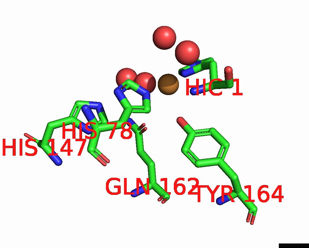
Mono view
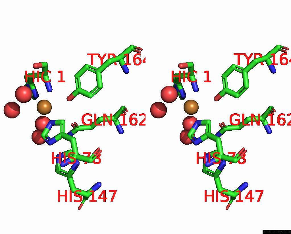
Stereo pair view
Mono view
Stereo pair view
A full contact list of Copper with other atoms in the Cu binding
site number 1 of X-Ray Structure of Lpmo at 1.45X10^6 Gy within 5.0Å range:
|
Reference:
T.Tandrup,
S.J.Muderspach,
S.Banerjee,
G.Santoni,
J.O.Ipsen,
C.Hernandez-Rollan,
M.H.H.Norholm,
K.S.Johansen,
F.Meilleur,
L.Lo Leggio.
Changes in Active-Site Geometry on X-Ray Photoreduction of A Lytic Polysaccharide Monooxygenase Active-Site Copper and Saccharide Binding. Iucrj V. 9 666 2022.
ISSN: ESSN 2052-2525
PubMed: 36071795
DOI: 10.1107/S2052252522007175
Page generated: Mon Jul 14 08:18:36 2025
ISSN: ESSN 2052-2525
PubMed: 36071795
DOI: 10.1107/S2052252522007175
Last articles
Fe in 2YXOFe in 2YRS
Fe in 2YXC
Fe in 2YNM
Fe in 2YVJ
Fe in 2YP1
Fe in 2YU2
Fe in 2YU1
Fe in 2YQB
Fe in 2YOO