Copper »
PDB 7o3e-7pyi »
7pxj »
Copper in PDB 7pxj: X-Ray Structure of Lpmo at 5.99X10^4 Gy
Enzymatic activity of X-Ray Structure of Lpmo at 5.99X10^4 Gy
All present enzymatic activity of X-Ray Structure of Lpmo at 5.99X10^4 Gy:
1.14.99.56;
1.14.99.56;
Protein crystallography data
The structure of X-Ray Structure of Lpmo at 5.99X10^4 Gy, PDB code: 7pxj
was solved by
T.Tandrup,
L.Lo Leggio,
with X-Ray Crystallography technique. A brief refinement statistics is given in the table below:
| Resolution Low / High (Å) | 44.23 / 1.75 |
| Space group | P 41 3 2 |
| Cell size a, b, c (Å), α, β, γ (°) | 124.99, 124.99, 124.99, 90, 90, 90 |
| R / Rfree (%) | 18.3 / 21.5 |
Copper Binding Sites:
The binding sites of Copper atom in the X-Ray Structure of Lpmo at 5.99X10^4 Gy
(pdb code 7pxj). This binding sites where shown within
5.0 Angstroms radius around Copper atom.
In total only one binding site of Copper was determined in the X-Ray Structure of Lpmo at 5.99X10^4 Gy, PDB code: 7pxj:
In total only one binding site of Copper was determined in the X-Ray Structure of Lpmo at 5.99X10^4 Gy, PDB code: 7pxj:
Copper binding site 1 out of 1 in 7pxj
Go back to
Copper binding site 1 out
of 1 in the X-Ray Structure of Lpmo at 5.99X10^4 Gy
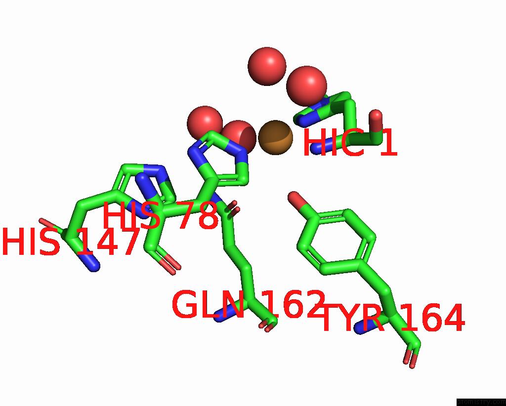
Mono view
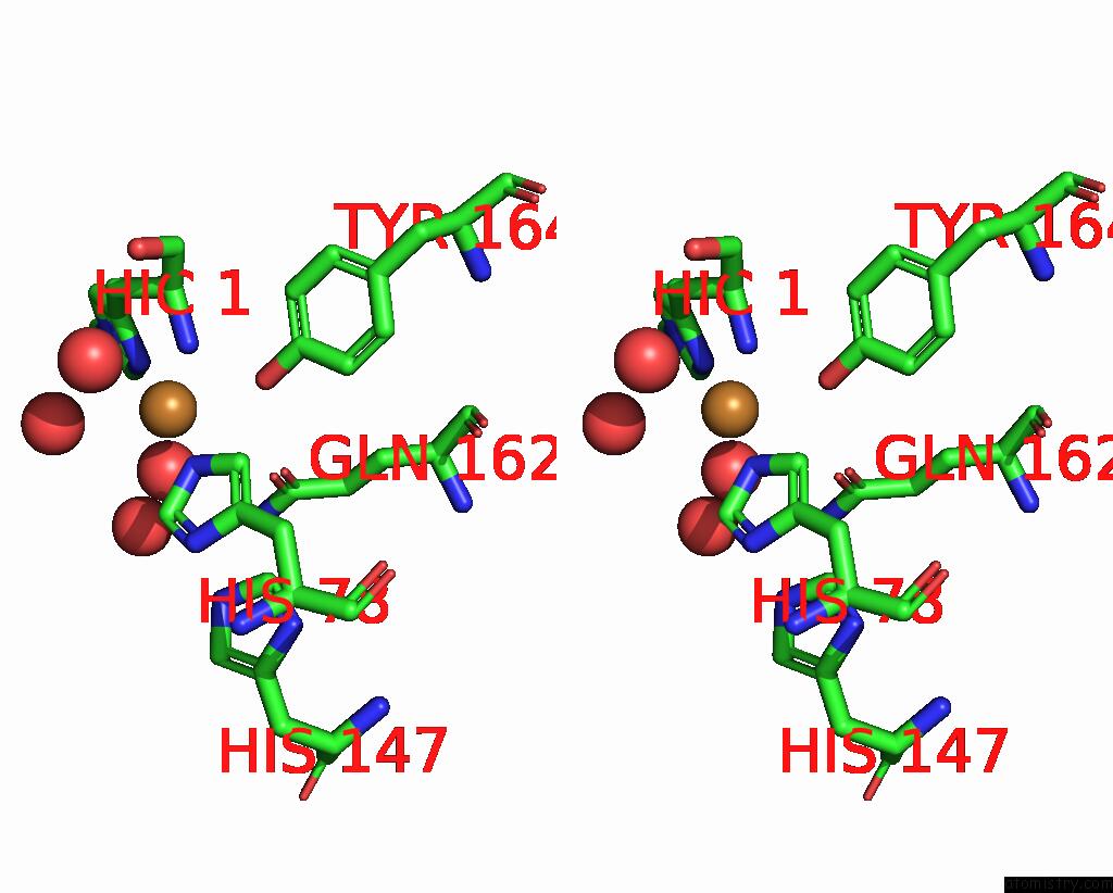
Stereo pair view
Mono view
Stereo pair view
A full contact list of Copper with other atoms in the Cu binding
site number 1 of X-Ray Structure of Lpmo at 5.99X10^4 Gy within 5.0Å range:
|
Reference:
T.Tandrup,
S.J.Muderspach,
S.Banerjee,
G.Santoni,
J.O.Ipsen,
C.Hernandez-Rollan,
M.H.H.Norholm,
K.S.Johansen,
F.Meilleur,
L.Lo Leggio.
Changes in Active-Site Geometry on X-Ray Photoreduction of A Lytic Polysaccharide Monooxygenase Active-Site Copper and Saccharide Binding. Iucrj V. 9 666 2022.
ISSN: ESSN 2052-2525
PubMed: 36071795
DOI: 10.1107/S2052252522007175
Page generated: Wed Jul 31 08:44:51 2024
ISSN: ESSN 2052-2525
PubMed: 36071795
DOI: 10.1107/S2052252522007175
Last articles
Zn in 9JYWZn in 9IR4
Zn in 9IR3
Zn in 9GMX
Zn in 9GMW
Zn in 9JEJ
Zn in 9ERF
Zn in 9ERE
Zn in 9EGV
Zn in 9EGW