Copper »
PDB 7ev9-7o3c »
7icj »
Copper in PDB 7icj: Dna Polymerase Beta (Pol B) (E.C.2.7.7.7) Complexed with Six Base Pairs of Dna; Soaked in the Presence of CUCL2 (0.1 Millimolar)
Protein crystallography data
The structure of Dna Polymerase Beta (Pol B) (E.C.2.7.7.7) Complexed with Six Base Pairs of Dna; Soaked in the Presence of CUCL2 (0.1 Millimolar), PDB code: 7icj
was solved by
H.Pelletier,
M.R.Sawaya,
with X-Ray Crystallography technique. A brief refinement statistics is given in the table below:
| Resolution Low / High (Å) | 20.00 / 3.50 |
| Space group | P 21 21 2 |
| Cell size a, b, c (Å), α, β, γ (°) | 177.010, 57.749, 48.582, 90.00, 90.00, 90.00 |
| R / Rfree (%) | 16.3 / n/a |
Other elements in 7icj:
The structure of Dna Polymerase Beta (Pol B) (E.C.2.7.7.7) Complexed with Six Base Pairs of Dna; Soaked in the Presence of CUCL2 (0.1 Millimolar) also contains other interesting chemical elements:
| Sodium | (Na) | 1 atom |
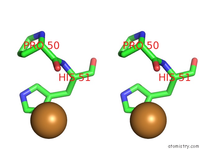
Copper Binding Sites:
The binding sites of Copper atom in the Dna Polymerase Beta (Pol B) (E.C.2.7.7.7) Complexed with Six Base Pairs of Dna; Soaked in the Presence of CUCL2 (0.1 Millimolar)
(pdb code 7icj). This binding sites where shown within
5.0 Angstroms radius around Copper atom.
In total only one binding site of Copper was determined in the Dna Polymerase Beta (Pol B) (E.C.2.7.7.7) Complexed with Six Base Pairs of Dna; Soaked in the Presence of CUCL2 (0.1 Millimolar), PDB code: 7icj:
In total only one binding site of Copper was determined in the Dna Polymerase Beta (Pol B) (E.C.2.7.7.7) Complexed with Six Base Pairs of Dna; Soaked in the Presence of CUCL2 (0.1 Millimolar), PDB code: 7icj:
Copper binding site 1 out of 1 in 7icj
Go back to
Copper binding site 1 out
of 1 in the Dna Polymerase Beta (Pol B) (E.C.2.7.7.7) Complexed with Six Base Pairs of Dna; Soaked in the Presence of CUCL2 (0.1 Millimolar)

Mono view
Stereo pair view
Mono view
Stereo pair view
A full contact list of Copper with other atoms in the Cu binding
site number 1 of Dna Polymerase Beta (Pol B) (E.C.2.7.7.7) Complexed with Six Base Pairs of Dna; Soaked in the Presence of CUCL2 (0.1 Millimolar) within 5.0Å range:
|
Reference:
H.Pelletier,
M.R.Sawaya,
W.Wolfle,
S.H.Wilson,
J.Kraut.
A Structural Basis For Metal Ion Mutagenicity and Nucleotide Selectivity in Human Dna Polymerase Beta Biochemistry V. 35 12762 1996.
ISSN: ISSN 0006-2960
PubMed: 8841119
DOI: 10.1021/BI9529566
Page generated: Mon Jul 14 08:03:14 2025
ISSN: ISSN 0006-2960
PubMed: 8841119
DOI: 10.1021/BI9529566
Last articles
Na in 7C1ZNa in 7C65
Na in 7C1Y
Na in 7C1X
Na in 7C0W
Na in 7BYF
Na in 7C0O
Na in 7BTK
Na in 7BRS
Na in 7C0K