Copper in PDB 7atn: Cytochrome C Oxidase Structure in R-State
Enzymatic activity of Cytochrome C Oxidase Structure in R-State
All present enzymatic activity of Cytochrome C Oxidase Structure in R-State:
7.1.1.9;
Other elements in 7atn:
The structure of Cytochrome C Oxidase Structure in R-State also contains other interesting chemical elements:
Copper Binding Sites:
The binding sites of Copper atom in the Cytochrome C Oxidase Structure in R-State
(pdb code 7atn). This binding sites where shown within
5.0 Angstroms radius around Copper atom.
In total 3 binding sites of Copper where determined in the
Cytochrome C Oxidase Structure in R-State, PDB code: 7atn:
Jump to Copper binding site number:
1;
2;
3;
Copper binding site 1 out
of 3 in 7atn
Go back to
Copper Binding Sites List in 7atn
Copper binding site 1 out
of 3 in the Cytochrome C Oxidase Structure in R-State
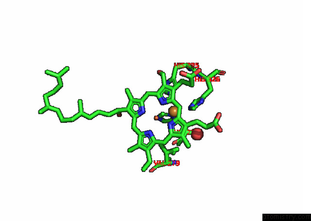
Mono view
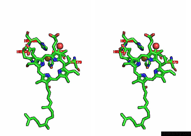
Stereo pair view
|
A full contact list of Copper with other atoms in the Cu binding
site number 1 of Cytochrome C Oxidase Structure in R-State within 5.0Å range:
probe
|
atom
|
residue
|
distance (Å)
|
B
|
Occ
|
A:Cu603
b:36.0
occ:1.00
|
ND1
|
A:HIS276
|
1.9
|
9.2
|
1.0
|
CE1
|
A:HIS276
|
2.2
|
9.2
|
1.0
|
NE2
|
A:HIS326
|
2.2
|
6.2
|
1.0
|
NE2
|
A:HIS325
|
2.5
|
7.1
|
1.0
|
CE1
|
A:HIS326
|
3.0
|
6.2
|
1.0
|
CG
|
A:HIS276
|
3.2
|
9.2
|
1.0
|
CD2
|
A:HIS326
|
3.3
|
6.2
|
1.0
|
CE1
|
A:HIS325
|
3.4
|
7.1
|
1.0
|
NE2
|
A:HIS276
|
3.5
|
9.2
|
1.0
|
CD2
|
A:HIS325
|
3.5
|
7.1
|
1.0
|
NA
|
A:HEA605
|
3.6
|
12.2
|
1.0
|
C1A
|
A:HEA605
|
3.8
|
12.2
|
1.0
|
FE
|
A:HEA605
|
3.9
|
12.2
|
1.0
|
CD2
|
A:HIS276
|
3.9
|
9.2
|
1.0
|
CB
|
A:HIS276
|
4.0
|
9.2
|
1.0
|
C4A
|
A:HEA605
|
4.1
|
12.2
|
1.0
|
ND1
|
A:HIS326
|
4.1
|
6.2
|
1.0
|
CHA
|
A:HEA605
|
4.2
|
12.2
|
1.0
|
ND
|
A:HEA605
|
4.2
|
12.2
|
1.0
|
CG
|
A:HIS326
|
4.3
|
6.2
|
1.0
|
C4D
|
A:HEA605
|
4.3
|
12.2
|
1.0
|
C2A
|
A:HEA605
|
4.4
|
12.2
|
1.0
|
CA
|
A:HIS276
|
4.4
|
9.2
|
1.0
|
C3A
|
A:HEA605
|
4.5
|
12.2
|
1.0
|
NB
|
A:HEA605
|
4.5
|
12.2
|
1.0
|
ND1
|
A:HIS325
|
4.6
|
7.1
|
1.0
|
O
|
A:HOH746
|
4.6
|
9.9
|
1.0
|
CHB
|
A:HEA605
|
4.6
|
12.2
|
1.0
|
CG
|
A:HIS325
|
4.7
|
7.1
|
1.0
|
CG2
|
A:VAL279
|
4.8
|
9.3
|
1.0
|
C1B
|
A:HEA605
|
4.8
|
12.2
|
1.0
|
NC
|
A:HEA605
|
5.0
|
12.2
|
1.0
|
|
Copper binding site 2 out
of 3 in 7atn
Go back to
Copper Binding Sites List in 7atn
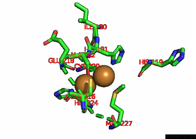
Copper binding site 2 out
of 3 in the Cytochrome C Oxidase Structure in R-State
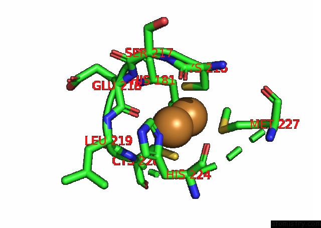
Mono view
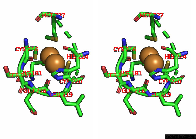
Stereo pair view
|
A full contact list of Copper with other atoms in the Cu binding
site number 2 of Cytochrome C Oxidase Structure in R-State within 5.0Å range:
probe
|
atom
|
residue
|
distance (Å)
|
B
|
Occ
|
B:Cu301
b:41.6
occ:1.00
|
CU1
|
B:CUA301
|
0.0
|
41.6
|
1.0
|
ND1
|
B:HIS224
|
2.1
|
8.2
|
1.0
|
O
|
B:GLU218
|
2.6
|
13.7
|
1.0
|
CU2
|
B:CUA301
|
2.7
|
41.6
|
1.0
|
SG
|
B:CYS220
|
2.8
|
10.6
|
1.0
|
CE1
|
B:HIS224
|
3.0
|
8.2
|
1.0
|
SG
|
B:CYS216
|
3.0
|
9.2
|
1.0
|
CG
|
B:HIS224
|
3.2
|
8.2
|
1.0
|
O
|
B:HIS224
|
3.4
|
8.2
|
1.0
|
CB
|
B:CYS220
|
3.4
|
10.6
|
1.0
|
CB
|
B:CYS216
|
3.5
|
9.2
|
1.0
|
CA
|
B:HIS224
|
3.5
|
8.2
|
1.0
|
CB
|
B:HIS224
|
3.6
|
8.2
|
1.0
|
N
|
B:CYS220
|
3.7
|
10.6
|
1.0
|
C
|
B:GLU218
|
3.7
|
13.7
|
1.0
|
C
|
B:HIS224
|
3.9
|
8.2
|
1.0
|
SD
|
B:MET227
|
4.1
|
6.8
|
1.0
|
NE2
|
B:HIS224
|
4.1
|
8.2
|
1.0
|
CA
|
B:CYS220
|
4.2
|
10.6
|
1.0
|
CD2
|
B:HIS224
|
4.2
|
8.2
|
1.0
|
C
|
B:LEU219
|
4.4
|
7.1
|
1.0
|
CA
|
B:LEU219
|
4.4
|
7.1
|
1.0
|
N
|
B:GLU218
|
4.5
|
13.7
|
1.0
|
N
|
B:LEU219
|
4.5
|
7.1
|
1.0
|
C
|
B:CYS216
|
4.5
|
9.2
|
1.0
|
CG
|
B:MET227
|
4.5
|
6.8
|
1.0
|
O
|
B:CYS216
|
4.6
|
9.2
|
1.0
|
CA
|
B:CYS216
|
4.6
|
9.2
|
1.0
|
ND1
|
B:HIS181
|
4.7
|
7.7
|
1.0
|
CA
|
B:GLU218
|
4.7
|
13.7
|
1.0
|
N
|
B:HIS224
|
4.8
|
8.2
|
1.0
|
N
|
B:SER217
|
4.9
|
10.3
|
1.0
|
|
Copper binding site 3 out
of 3 in 7atn
Go back to
Copper Binding Sites List in 7atn
Copper binding site 3 out
of 3 in the Cytochrome C Oxidase Structure in R-State
Mono view
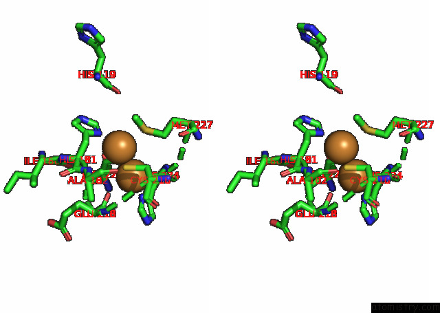
Stereo pair view
|
A full contact list of Copper with other atoms in the Cu binding
site number 3 of Cytochrome C Oxidase Structure in R-State within 5.0Å range:
probe
|
atom
|
residue
|
distance (Å)
|
B
|
Occ
|
B:Cu301
b:41.6
occ:1.00
|
CU2
|
B:CUA301
|
0.0
|
41.6
|
1.0
|
ND1
|
B:HIS181
|
2.1
|
7.7
|
1.0
|
SD
|
B:MET227
|
2.6
|
6.8
|
1.0
|
CU1
|
B:CUA301
|
2.7
|
41.6
|
1.0
|
SG
|
B:CYS216
|
2.8
|
9.2
|
1.0
|
SG
|
B:CYS220
|
3.0
|
10.6
|
1.0
|
CE
|
B:MET227
|
3.0
|
6.8
|
1.0
|
CE1
|
B:HIS181
|
3.1
|
7.7
|
1.0
|
CG
|
B:HIS181
|
3.1
|
7.7
|
1.0
|
CB
|
B:CYS220
|
3.3
|
10.6
|
1.0
|
CB
|
B:HIS181
|
3.4
|
7.7
|
1.0
|
CB
|
B:CYS216
|
3.8
|
9.2
|
1.0
|
CG
|
B:MET227
|
3.9
|
6.8
|
1.0
|
CA
|
B:HIS181
|
4.0
|
7.7
|
1.0
|
O
|
B:GLU218
|
4.1
|
13.7
|
1.0
|
NE2
|
B:HIS181
|
4.2
|
7.7
|
1.0
|
CD2
|
B:HIS181
|
4.3
|
7.7
|
1.0
|
O
|
B:ILE180
|
4.6
|
6.9
|
1.0
|
CA
|
B:CYS220
|
4.8
|
10.6
|
1.0
|
ND1
|
B:HIS224
|
4.8
|
8.2
|
1.0
|
O
|
B:HIS119
|
4.9
|
9.1
|
1.0
|
N
|
B:ALA182
|
4.9
|
9.2
|
1.0
|
N
|
B:HIS181
|
5.0
|
7.7
|
1.0
|
|
Reference:
F.Kolbe,
S.Safarian,
H.Michel.
Cytochrome C Oxidase Structure in R-State To Be Published.
Page generated: Wed Jul 31 08:19:46 2024
|