Copper »
PDB 6y6y-6zut »
6yde »
Copper in PDB 6yde: X-Ray Structure of Lpmo
Protein crystallography data
The structure of X-Ray Structure of Lpmo, PDB code: 6yde
was solved by
T.Tandrup,
T.Tryfona,
K.E.H.Frandsen,
K.S.Johansen,
P.Dupree,
L.Lo Leggio,
with X-Ray Crystallography technique. A brief refinement statistics is given in the table below:
| Resolution Low / High (Å) | 50.00 / 2.20 |
| Space group | P 21 21 2 |
| Cell size a, b, c (Å), α, β, γ (°) | 39.550, 124.370, 51.670, 90.00, 90.00, 90.00 |
| R / Rfree (%) | 19.4 / 26.4 |
Copper Binding Sites:
The binding sites of Copper atom in the X-Ray Structure of Lpmo
(pdb code 6yde). This binding sites where shown within
5.0 Angstroms radius around Copper atom.
In total only one binding site of Copper was determined in the X-Ray Structure of Lpmo, PDB code: 6yde:
In total only one binding site of Copper was determined in the X-Ray Structure of Lpmo, PDB code: 6yde:
Copper binding site 1 out of 1 in 6yde
Go back to
Copper binding site 1 out
of 1 in the X-Ray Structure of Lpmo
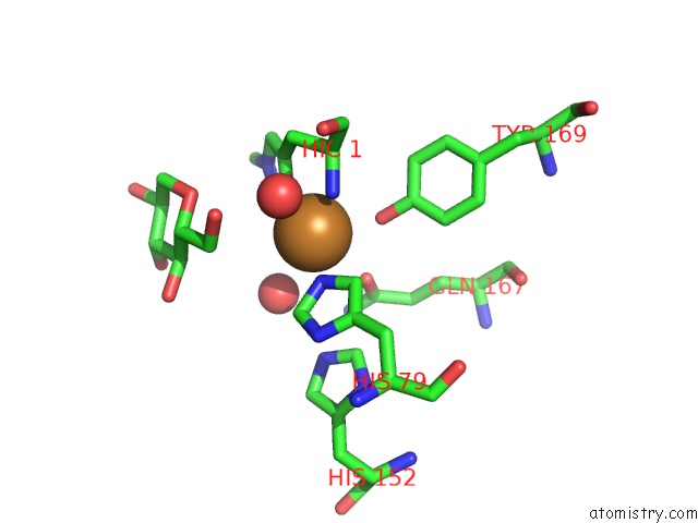
Mono view
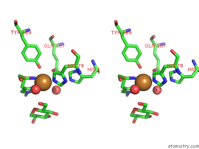
Stereo pair view
Mono view
Stereo pair view
A full contact list of Copper with other atoms in the Cu binding
site number 1 of X-Ray Structure of Lpmo within 5.0Å range:
|
Reference:
T.Tandrup,
T.Tryfona,
K.E.H.Frandsen,
K.S.Johansen,
P.Dupree,
L.Lo Leggio.
Oligosaccharide Binding and Thermostability of Two Related AA9 Lytic Polysaccharide Monooxygenases. Biochemistry V. 59 3347 2020.
ISSN: ISSN 0006-2960
PubMed: 32818374
DOI: 10.1021/ACS.BIOCHEM.0C00312
Page generated: Wed Jul 31 07:53:41 2024
ISSN: ISSN 0006-2960
PubMed: 32818374
DOI: 10.1021/ACS.BIOCHEM.0C00312
Last articles
Zn in 9JYWZn in 9IR4
Zn in 9IR3
Zn in 9GMX
Zn in 9GMW
Zn in 9JEJ
Zn in 9ERF
Zn in 9ERE
Zn in 9EGV
Zn in 9EGW