Copper »
PDB 6l8s-6pvy »
6paz »
Copper in PDB 6paz: Oxidized Mutant P80I Pseudoazurin From A. Faecalis
Protein crystallography data
The structure of Oxidized Mutant P80I Pseudoazurin From A. Faecalis, PDB code: 6paz
was solved by
E.T.Adman,
C.A.P.Libeu,
with X-Ray Crystallography technique. A brief refinement statistics is given in the table below:
| Resolution Low / High (Å) | 18.26 / 1.91 |
| Space group | P 65 |
| Cell size a, b, c (Å), α, β, γ (°) | 50.790, 50.790, 98.250, 90.00, 90.00, 120.00 |
| R / Rfree (%) | 19 / n/a |
Copper Binding Sites:
The binding sites of Copper atom in the Oxidized Mutant P80I Pseudoazurin From A. Faecalis
(pdb code 6paz). This binding sites where shown within
5.0 Angstroms radius around Copper atom.
In total only one binding site of Copper was determined in the Oxidized Mutant P80I Pseudoazurin From A. Faecalis, PDB code: 6paz:
In total only one binding site of Copper was determined in the Oxidized Mutant P80I Pseudoazurin From A. Faecalis, PDB code: 6paz:
Copper binding site 1 out of 1 in 6paz
Go back to
Copper binding site 1 out
of 1 in the Oxidized Mutant P80I Pseudoazurin From A. Faecalis
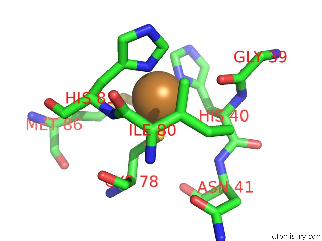
Mono view
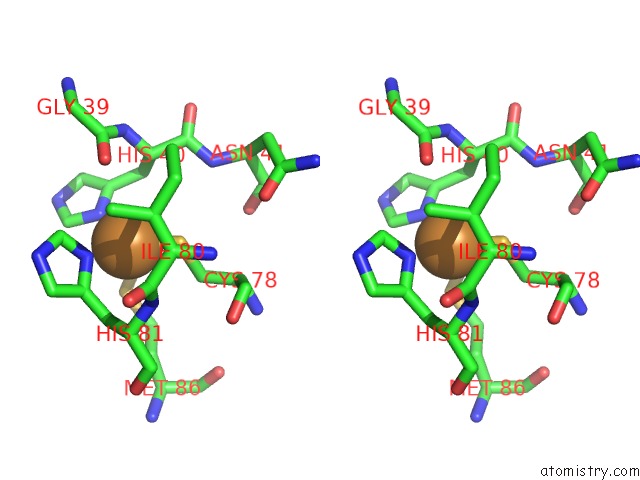
Stereo pair view
Mono view
Stereo pair view
A full contact list of Copper with other atoms in the Cu binding
site number 1 of Oxidized Mutant P80I Pseudoazurin From A. Faecalis within 5.0Å range:
|
Reference:
C.A.Libeu,
M.Kukimoto,
M.Nishiyama,
S.Horinouchi,
E.T.Adman.
Site-Directed Mutants of Pseudoazurin: Explanation of Increased Redox Potentials From X-Ray Structures and From Calculation of Redox Potential Differences. Biochemistry V. 36 13160 1997.
ISSN: ISSN 0006-2960
PubMed: 9341204
DOI: 10.1021/BI9704111
Page generated: Mon Jul 14 06:36:43 2025
ISSN: ISSN 0006-2960
PubMed: 9341204
DOI: 10.1021/BI9704111
Last articles
Fe in 2YXOFe in 2YRS
Fe in 2YXC
Fe in 2YNM
Fe in 2YVJ
Fe in 2YP1
Fe in 2YU2
Fe in 2YU1
Fe in 2YQB
Fe in 2YOO