Copper »
PDB 6ied-6l58 »
6koc »
Copper in PDB 6koc: X-Ray Structure of the Proton-Pumping Cytochrome AA3-600 Menaquinol Oxidase From Bacillus Subtilis Complexed with 3-Iodo-N-Oxo-2-Heptyl- 4-Hydroxyquinoline
Protein crystallography data
The structure of X-Ray Structure of the Proton-Pumping Cytochrome AA3-600 Menaquinol Oxidase From Bacillus Subtilis Complexed with 3-Iodo-N-Oxo-2-Heptyl- 4-Hydroxyquinoline, PDB code: 6koc
was solved by
J.Xu,
Z.Ding,
B.Liu,
J.Li,
R.B.Gennis,
J.Zhu,
with X-Ray Crystallography technique. A brief refinement statistics is given in the table below:
| Resolution Low / High (Å) | 48.27 / 3.80 |
| Space group | P 1 21 1 |
| Cell size a, b, c (Å), α, β, γ (°) | 113.930, 162.200, 151.320, 90.00, 109.76, 90.00 |
| R / Rfree (%) | 32.5 / 36.9 |
Other elements in 6koc:
The structure of X-Ray Structure of the Proton-Pumping Cytochrome AA3-600 Menaquinol Oxidase From Bacillus Subtilis Complexed with 3-Iodo-N-Oxo-2-Heptyl- 4-Hydroxyquinoline also contains other interesting chemical elements:
| Iodine | (I) | 2 atoms |
| Iron | (Fe) | 4 atoms |
Copper Binding Sites:
The binding sites of Copper atom in the X-Ray Structure of the Proton-Pumping Cytochrome AA3-600 Menaquinol Oxidase From Bacillus Subtilis Complexed with 3-Iodo-N-Oxo-2-Heptyl- 4-Hydroxyquinoline
(pdb code 6koc). This binding sites where shown within
5.0 Angstroms radius around Copper atom.
In total 2 binding sites of Copper where determined in the X-Ray Structure of the Proton-Pumping Cytochrome AA3-600 Menaquinol Oxidase From Bacillus Subtilis Complexed with 3-Iodo-N-Oxo-2-Heptyl- 4-Hydroxyquinoline, PDB code: 6koc:
Jump to Copper binding site number: 1; 2;
In total 2 binding sites of Copper where determined in the X-Ray Structure of the Proton-Pumping Cytochrome AA3-600 Menaquinol Oxidase From Bacillus Subtilis Complexed with 3-Iodo-N-Oxo-2-Heptyl- 4-Hydroxyquinoline, PDB code: 6koc:
Jump to Copper binding site number: 1; 2;
Copper binding site 1 out of 2 in 6koc
Go back to
Copper binding site 1 out
of 2 in the X-Ray Structure of the Proton-Pumping Cytochrome AA3-600 Menaquinol Oxidase From Bacillus Subtilis Complexed with 3-Iodo-N-Oxo-2-Heptyl- 4-Hydroxyquinoline
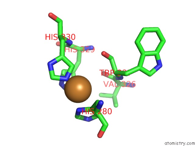
Mono view
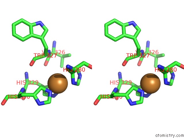
Stereo pair view
Mono view
Stereo pair view
A full contact list of Copper with other atoms in the Cu binding
site number 1 of X-Ray Structure of the Proton-Pumping Cytochrome AA3-600 Menaquinol Oxidase From Bacillus Subtilis Complexed with 3-Iodo-N-Oxo-2-Heptyl- 4-Hydroxyquinoline within 5.0Å range:
|
Copper binding site 2 out of 2 in 6koc
Go back to
Copper binding site 2 out
of 2 in the X-Ray Structure of the Proton-Pumping Cytochrome AA3-600 Menaquinol Oxidase From Bacillus Subtilis Complexed with 3-Iodo-N-Oxo-2-Heptyl- 4-Hydroxyquinoline
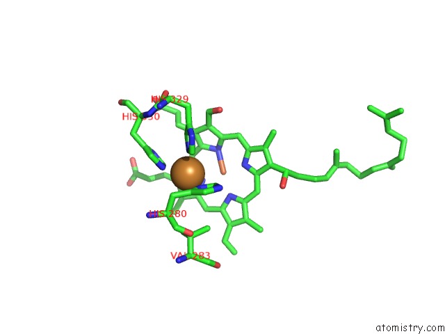
Mono view
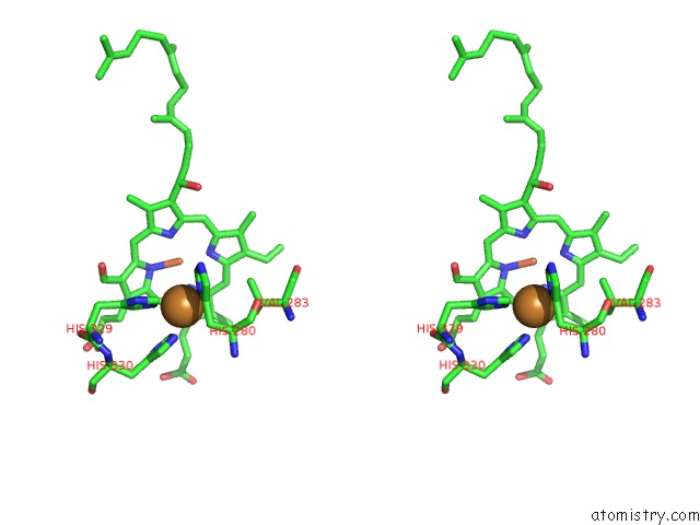
Stereo pair view
Mono view
Stereo pair view
A full contact list of Copper with other atoms in the Cu binding
site number 2 of X-Ray Structure of the Proton-Pumping Cytochrome AA3-600 Menaquinol Oxidase From Bacillus Subtilis Complexed with 3-Iodo-N-Oxo-2-Heptyl- 4-Hydroxyquinoline within 5.0Å range:
|
Reference:
J.Xu,
Z.Ding,
B.Liu,
S.M.Yi,
J.Li,
Z.Zhang,
Y.Liu,
J.Li,
L.Liu,
A.Zhou,
R.B.Gennis,
J.Zhu.
Structure of the Cytochrome AA3-600 Heme-Copper Menaquinol Oxidase Bound to Inhibitor Hqno Shows TM0 Is Part of the Quinol Binding Site Proc.Natl.Acad.Sci.Usa 2010.
ISSN: ESSN 1091-6490
DOI: 10.1073/PNAS.1915013117
Page generated: Wed Jul 31 06:28:45 2024
ISSN: ESSN 1091-6490
DOI: 10.1073/PNAS.1915013117
Last articles
Zn in 9MJ5Zn in 9HNW
Zn in 9G0L
Zn in 9FNE
Zn in 9DZN
Zn in 9E0I
Zn in 9D32
Zn in 9DAK
Zn in 8ZXC
Zn in 8ZUF