Copper »
PDB 5wbd-5z85 »
5xmo »
Copper in PDB 5xmo: X-Ray Crystal Structure of Pseudoazurin MET16PHE/THR36LYS Variant
Protein crystallography data
The structure of X-Ray Crystal Structure of Pseudoazurin MET16PHE/THR36LYS Variant, PDB code: 5xmo
was solved by
T.Yamaguchi,
T.Kohzuma,
with X-Ray Crystallography technique. A brief refinement statistics is given in the table below:
| Resolution Low / High (Å) | 46.05 / 1.19 |
| Space group | C 1 2 1 |
| Cell size a, b, c (Å), α, β, γ (°) | 93.170, 41.160, 34.830, 90.00, 98.71, 90.00 |
| R / Rfree (%) | 12.8 / 16.1 |
Copper Binding Sites:
The binding sites of Copper atom in the X-Ray Crystal Structure of Pseudoazurin MET16PHE/THR36LYS Variant
(pdb code 5xmo). This binding sites where shown within
5.0 Angstroms radius around Copper atom.
In total 2 binding sites of Copper where determined in the X-Ray Crystal Structure of Pseudoazurin MET16PHE/THR36LYS Variant, PDB code: 5xmo:
Jump to Copper binding site number: 1; 2;
In total 2 binding sites of Copper where determined in the X-Ray Crystal Structure of Pseudoazurin MET16PHE/THR36LYS Variant, PDB code: 5xmo:
Jump to Copper binding site number: 1; 2;
Copper binding site 1 out of 2 in 5xmo
Go back to
Copper binding site 1 out
of 2 in the X-Ray Crystal Structure of Pseudoazurin MET16PHE/THR36LYS Variant
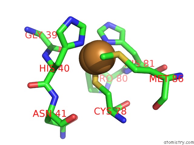
Mono view
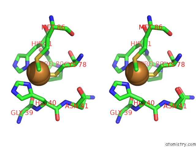
Stereo pair view
Mono view
Stereo pair view
A full contact list of Copper with other atoms in the Cu binding
site number 1 of X-Ray Crystal Structure of Pseudoazurin MET16PHE/THR36LYS Variant within 5.0Å range:
|
Copper binding site 2 out of 2 in 5xmo
Go back to
Copper binding site 2 out
of 2 in the X-Ray Crystal Structure of Pseudoazurin MET16PHE/THR36LYS Variant
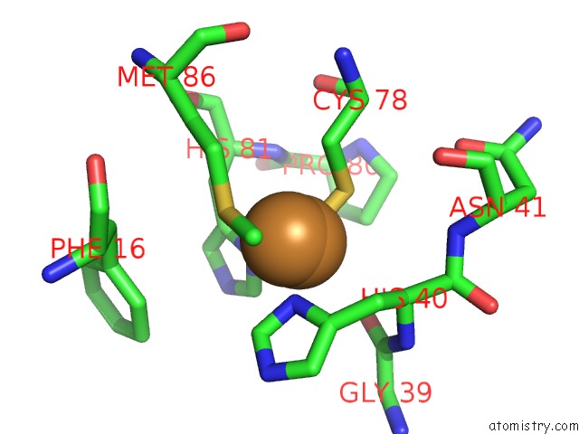
Mono view
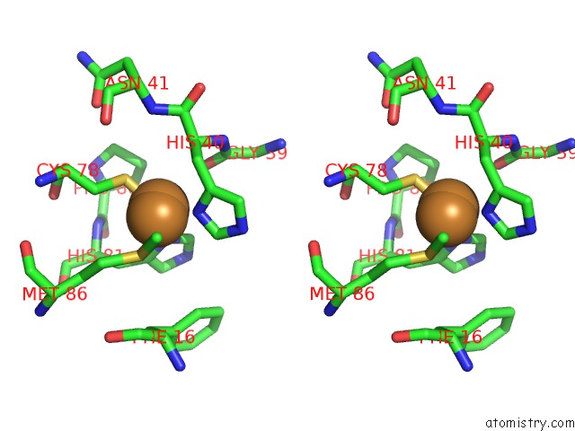
Stereo pair view
Mono view
Stereo pair view
A full contact list of Copper with other atoms in the Cu binding
site number 2 of X-Ray Crystal Structure of Pseudoazurin MET16PHE/THR36LYS Variant within 5.0Å range:
|
Reference:
T.Yamaguchi,
N.Takebayashi,
K.Akao,
C.Sakai,
T.Kohzuma.
X-Ray Crystallographic Analysis of M16F/T36K Double Mutant of Pseudoazurin Photon Factory Activity V. 34 2017REPORT.
ISSN: ISSN 0912-1803
Page generated: Wed Jul 31 05:22:51 2024
ISSN: ISSN 0912-1803
Last articles
Zn in 9JYWZn in 9IR4
Zn in 9IR3
Zn in 9GMX
Zn in 9GMW
Zn in 9JEJ
Zn in 9ERF
Zn in 9ERE
Zn in 9EGV
Zn in 9EGW