Copper »
PDB 5onx-5wbc »
5wau »
Copper in PDB 5wau: Crystal Structure of Co-Bound Cytochrome C Oxidase Determined By Synchrotron X-Ray Crystallography at 100 K
Enzymatic activity of Crystal Structure of Co-Bound Cytochrome C Oxidase Determined By Synchrotron X-Ray Crystallography at 100 K
All present enzymatic activity of Crystal Structure of Co-Bound Cytochrome C Oxidase Determined By Synchrotron X-Ray Crystallography at 100 K:
1.9.3.1;
1.9.3.1;
Protein crystallography data
The structure of Crystal Structure of Co-Bound Cytochrome C Oxidase Determined By Synchrotron X-Ray Crystallography at 100 K, PDB code: 5wau
was solved by
R.Fromme,
I.Ishigami,
S.Y.Yeh,
N.Zatsepin,
T.Grant,
P.Fromme,
D.Rousseau,
with X-Ray Crystallography technique. A brief refinement statistics is given in the table below:
| Resolution Low / High (Å) | 39.00 / 1.95 |
| Space group | P 21 21 21 |
| Cell size a, b, c (Å), α, β, γ (°) | 177.923, 182.555, 208.450, 90.00, 90.00, 90.00 |
| R / Rfree (%) | 21.7 / 24.4 |
Other elements in 5wau:
The structure of Crystal Structure of Co-Bound Cytochrome C Oxidase Determined By Synchrotron X-Ray Crystallography at 100 K also contains other interesting chemical elements:
| Magnesium | (Mg) | 2 atoms |
| Zinc | (Zn) | 2 atoms |
| Iron | (Fe) | 4 atoms |
| Sodium | (Na) | 2 atoms |
Copper Binding Sites:
The binding sites of Copper atom in the Crystal Structure of Co-Bound Cytochrome C Oxidase Determined By Synchrotron X-Ray Crystallography at 100 K
(pdb code 5wau). This binding sites where shown within
5.0 Angstroms radius around Copper atom.
In total 6 binding sites of Copper where determined in the Crystal Structure of Co-Bound Cytochrome C Oxidase Determined By Synchrotron X-Ray Crystallography at 100 K, PDB code: 5wau:
Jump to Copper binding site number: 1; 2; 3; 4; 5; 6;
In total 6 binding sites of Copper where determined in the Crystal Structure of Co-Bound Cytochrome C Oxidase Determined By Synchrotron X-Ray Crystallography at 100 K, PDB code: 5wau:
Jump to Copper binding site number: 1; 2; 3; 4; 5; 6;
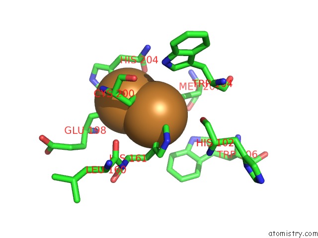
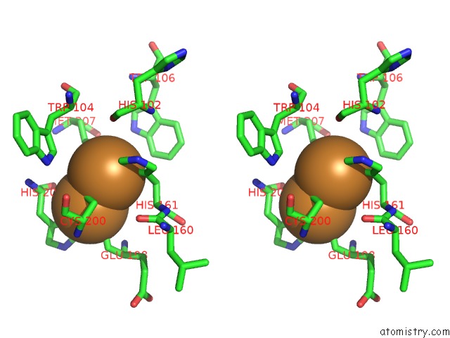
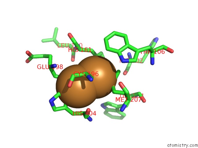
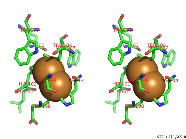
Copper binding site 1 out of 6 in 5wau
Go back to
Copper binding site 1 out
of 6 in the Crystal Structure of Co-Bound Cytochrome C Oxidase Determined By Synchrotron X-Ray Crystallography at 100 K
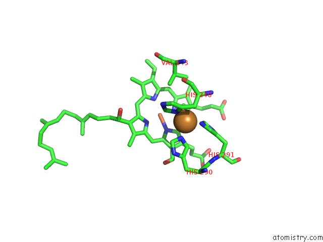
Mono view
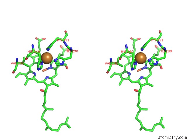
Stereo pair view
Mono view
Stereo pair view
A full contact list of Copper with other atoms in the Cu binding
site number 1 of Crystal Structure of Co-Bound Cytochrome C Oxidase Determined By Synchrotron X-Ray Crystallography at 100 K within 5.0Å range:
|
Copper binding site 2 out of 6 in 5wau
Go back to
Copper binding site 2 out
of 6 in the Crystal Structure of Co-Bound Cytochrome C Oxidase Determined By Synchrotron X-Ray Crystallography at 100 K
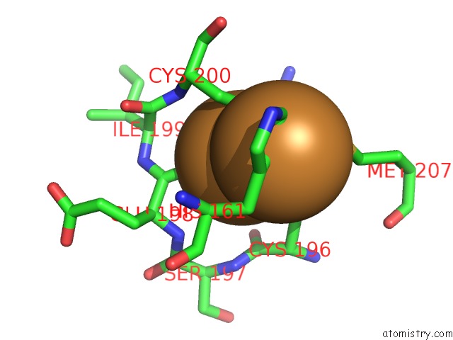
Mono view
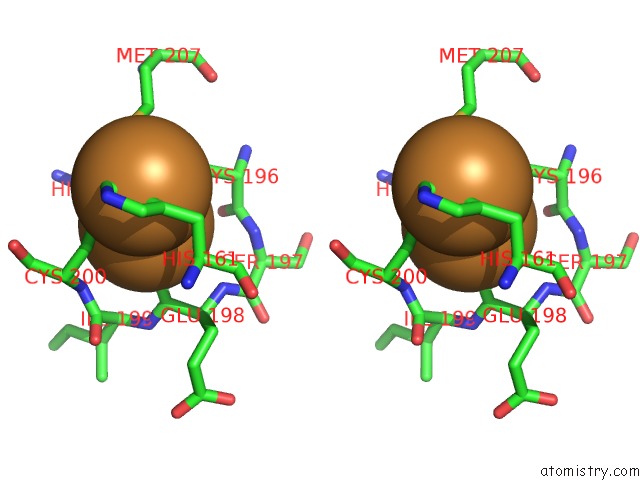
Stereo pair view
Mono view
Stereo pair view
A full contact list of Copper with other atoms in the Cu binding
site number 2 of Crystal Structure of Co-Bound Cytochrome C Oxidase Determined By Synchrotron X-Ray Crystallography at 100 K within 5.0Å range:
|
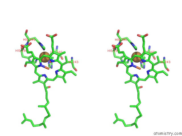
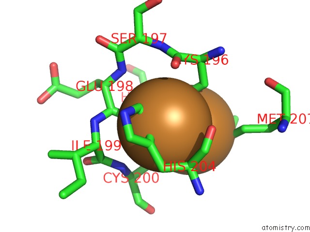
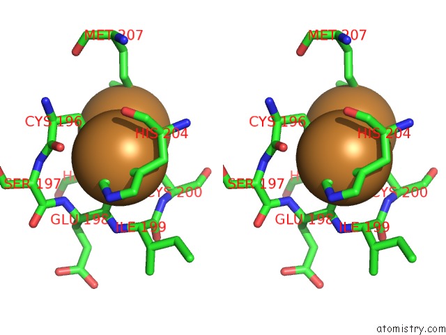
Copper binding site 3 out of 6 in 5wau
Go back to
Copper binding site 3 out
of 6 in the Crystal Structure of Co-Bound Cytochrome C Oxidase Determined By Synchrotron X-Ray Crystallography at 100 K
Mono view
Stereo pair view
Mono view
Stereo pair view
A full contact list of Copper with other atoms in the Cu binding
site number 3 of Crystal Structure of Co-Bound Cytochrome C Oxidase Determined By Synchrotron X-Ray Crystallography at 100 K within 5.0Å range:
|
Copper binding site 4 out of 6 in 5wau
Go back to
Copper binding site 4 out
of 6 in the Crystal Structure of Co-Bound Cytochrome C Oxidase Determined By Synchrotron X-Ray Crystallography at 100 K

Mono view
Stereo pair view
Mono view
Stereo pair view
A full contact list of Copper with other atoms in the Cu binding
site number 4 of Crystal Structure of Co-Bound Cytochrome C Oxidase Determined By Synchrotron X-Ray Crystallography at 100 K within 5.0Å range:
|
Copper binding site 5 out of 6 in 5wau
Go back to
Copper binding site 5 out
of 6 in the Crystal Structure of Co-Bound Cytochrome C Oxidase Determined By Synchrotron X-Ray Crystallography at 100 K
Mono view
Stereo pair view
Mono view
Stereo pair view
A full contact list of Copper with other atoms in the Cu binding
site number 5 of Crystal Structure of Co-Bound Cytochrome C Oxidase Determined By Synchrotron X-Ray Crystallography at 100 K within 5.0Å range:
|
Copper binding site 6 out of 6 in 5wau
Go back to
Copper binding site 6 out
of 6 in the Crystal Structure of Co-Bound Cytochrome C Oxidase Determined By Synchrotron X-Ray Crystallography at 100 K
Mono view
Stereo pair view
Mono view
Stereo pair view
A full contact list of Copper with other atoms in the Cu binding
site number 6 of Crystal Structure of Co-Bound Cytochrome C Oxidase Determined By Synchrotron X-Ray Crystallography at 100 K within 5.0Å range:
|
Reference:
I.Ishigami,
N.A.Zatsepin,
M.Hikita,
C.E.Conrad,
G.Nelson,
J.D.Coe,
S.Basu,
T.D.Grant,
M.H.Seaberg,
R.G.Sierra,
M.S.Hunter,
P.Fromme,
R.Fromme,
S.R.Yeh,
D.L.Rousseau.
Crystal Structure of Co-Bound Cytochrome C Oxidase Determined By Serial Femtosecond X-Ray Crystallography at Room Temperature. Proc. Natl. Acad. Sci. V. 114 8011 2017U.S.A..
ISSN: ESSN 1091-6490
PubMed: 28698372
DOI: 10.1073/PNAS.1705628114
Page generated: Wed Jul 31 05:17:33 2024
ISSN: ESSN 1091-6490
PubMed: 28698372
DOI: 10.1073/PNAS.1705628114
Last articles
Cl in 3FYOCl in 3FXZ
Cl in 3FXV
Cl in 3FXU
Cl in 3FWQ
Cl in 3FXQ
Cl in 3FW4
Cl in 3FWK
Cl in 3FWH
Cl in 3FW5