Copper »
PDB 5lww-5nlo »
5n05 »
Copper in PDB 5n05: X-Ray Crystal Structure of An Lpmo
Protein crystallography data
The structure of X-Ray Crystal Structure of An Lpmo, PDB code: 5n05
was solved by
K.E.H.Frandsen,
J.-C.N.Poulsen,
L.Lo Leggio,
with X-Ray Crystallography technique. A brief refinement statistics is given in the table below:
| Resolution Low / High (Å) | 44.07 / 1.58 |
| Space group | P 41 3 2 |
| Cell size a, b, c (Å), α, β, γ (°) | 124.640, 124.640, 124.640, 90.00, 90.00, 90.00 |
| R / Rfree (%) | 17.6 / 19.7 |
Other elements in 5n05:
The structure of X-Ray Crystal Structure of An Lpmo also contains other interesting chemical elements:
| Chlorine | (Cl) | 7 atoms |
Copper Binding Sites:
The binding sites of Copper atom in the X-Ray Crystal Structure of An Lpmo
(pdb code 5n05). This binding sites where shown within
5.0 Angstroms radius around Copper atom.
In total only one binding site of Copper was determined in the X-Ray Crystal Structure of An Lpmo, PDB code: 5n05:
In total only one binding site of Copper was determined in the X-Ray Crystal Structure of An Lpmo, PDB code: 5n05:
Copper binding site 1 out of 1 in 5n05
Go back to
Copper binding site 1 out
of 1 in the X-Ray Crystal Structure of An Lpmo
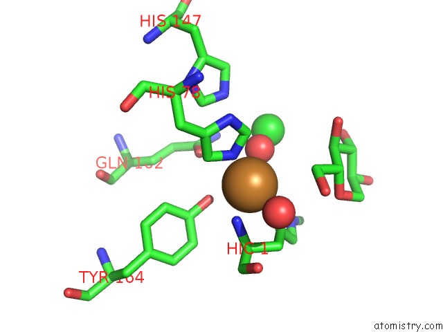
Mono view
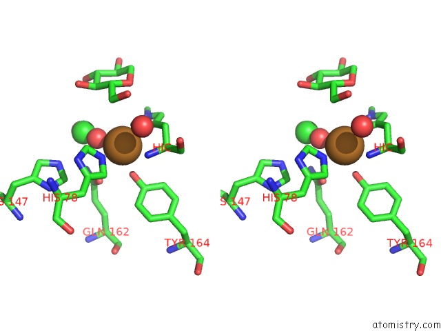
Stereo pair view
Mono view
Stereo pair view
A full contact list of Copper with other atoms in the Cu binding
site number 1 of X-Ray Crystal Structure of An Lpmo within 5.0Å range:
|
Reference:
K.E.H.Frandsen,
J.N.Poulsen,
T.Tandrup,
L.Lo Leggio.
Unliganded and Substrate Bound Structures of the Cellooligosaccharide Active Lytic Polysaccharide Monooxygenase LSAA9A at Low pH. Carbohydr. Res. V. 448 187 2017.
ISSN: ISSN 1873-426X
PubMed: 28364950
DOI: 10.1016/J.CARRES.2017.03.010
Page generated: Wed Jul 31 04:41:54 2024
ISSN: ISSN 1873-426X
PubMed: 28364950
DOI: 10.1016/J.CARRES.2017.03.010
Last articles
Zn in 9JYWZn in 9IR4
Zn in 9IR3
Zn in 9GMX
Zn in 9GMW
Zn in 9JEJ
Zn in 9ERF
Zn in 9ERE
Zn in 9EGV
Zn in 9EGW