Copper »
PDB 5ce9-5i0y »
5hr0 »
Copper in PDB 5hr0: Crystal Structure of Thioredoxin E101G Mutant
Protein crystallography data
The structure of Crystal Structure of Thioredoxin E101G Mutant, PDB code: 5hr0
was solved by
M.E.Noguera,
D.S.Vazquez,
E.I.Howard,
A.Cousido-Siah,
A.Mitschler,
A.Podjarny,
J.Santos,
with X-Ray Crystallography technique. A brief refinement statistics is given in the table below:
| Resolution Low / High (Å) | 19.86 / 1.31 |
| Space group | P 31 2 1 |
| Cell size a, b, c (Å), α, β, γ (°) | 50.043, 50.043, 163.291, 90.00, 90.00, 120.00 |
| R / Rfree (%) | 17.4 / 20.9 |
Copper Binding Sites:
The binding sites of Copper atom in the Crystal Structure of Thioredoxin E101G Mutant
(pdb code 5hr0). This binding sites where shown within
5.0 Angstroms radius around Copper atom.
In total 4 binding sites of Copper where determined in the Crystal Structure of Thioredoxin E101G Mutant, PDB code: 5hr0:
Jump to Copper binding site number: 1; 2; 3; 4;
In total 4 binding sites of Copper where determined in the Crystal Structure of Thioredoxin E101G Mutant, PDB code: 5hr0:
Jump to Copper binding site number: 1; 2; 3; 4;
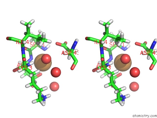
Copper binding site 1 out of 4 in 5hr0
Go back to
Copper binding site 1 out
of 4 in the Crystal Structure of Thioredoxin E101G Mutant

Mono view
Stereo pair view
Mono view
Stereo pair view
A full contact list of Copper with other atoms in the Cu binding
site number 1 of Crystal Structure of Thioredoxin E101G Mutant within 5.0Å range:
|
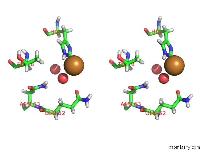
Copper binding site 2 out of 4 in 5hr0
Go back to
Copper binding site 2 out
of 4 in the Crystal Structure of Thioredoxin E101G Mutant

Mono view
Stereo pair view
Mono view
Stereo pair view
A full contact list of Copper with other atoms in the Cu binding
site number 2 of Crystal Structure of Thioredoxin E101G Mutant within 5.0Å range:
|
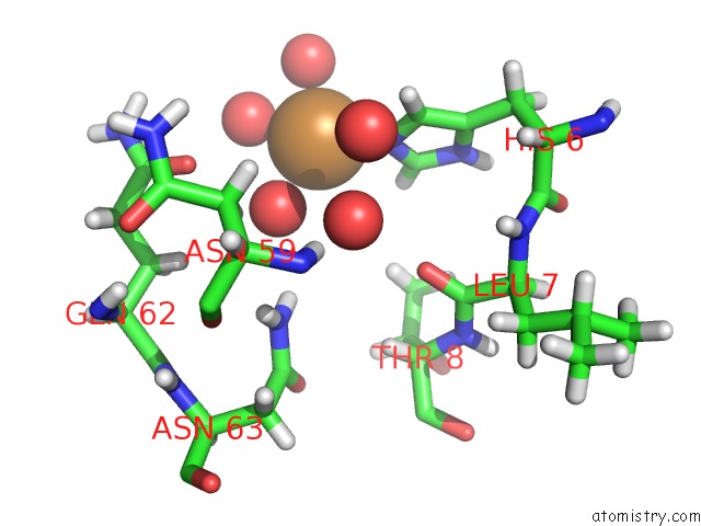
Copper binding site 3 out of 4 in 5hr0
Go back to
Copper binding site 3 out
of 4 in the Crystal Structure of Thioredoxin E101G Mutant

Mono view

Stereo pair view
Mono view
Stereo pair view
A full contact list of Copper with other atoms in the Cu binding
site number 3 of Crystal Structure of Thioredoxin E101G Mutant within 5.0Å range:
|
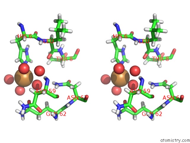
Copper binding site 4 out of 4 in 5hr0
Go back to
Copper binding site 4 out
of 4 in the Crystal Structure of Thioredoxin E101G Mutant
Mono view
Stereo pair view
Mono view
Stereo pair view
A full contact list of Copper with other atoms in the Cu binding
site number 4 of Crystal Structure of Thioredoxin E101G Mutant within 5.0Å range:
|
Reference:
M.E.Noguera,
D.S.Vazquez,
G.Ferrer-Sueta,
W.A.Agudelo,
E.Howard,
R.M.Rasia,
B.Manta,
A.Cousido-Siah,
A.Mitschler,
A.Podjarny,
J.Santos.
Structural Variability of E. Coli Thioredoxin Captured in the Crystal Structures of Single-Point Mutants. Sci Rep V. 7 42343 2017.
ISSN: ESSN 2045-2322
PubMed: 28181556
DOI: 10.1038/SREP42343
Page generated: Wed Jul 31 04:09:51 2024
ISSN: ESSN 2045-2322
PubMed: 28181556
DOI: 10.1038/SREP42343
Last articles
Cl in 5R9PCl in 5R9Q
Cl in 5R9O
Cl in 5R9N
Cl in 5R9M
Cl in 5R9K
Cl in 5R9L
Cl in 5R9J
Cl in 5R9I
Cl in 5R9G