Copper »
PDB 4ysu-5c92 »
5aci »
Copper in PDB 5aci: X-Ray Structure of Lpmo
Protein crystallography data
The structure of X-Ray Structure of Lpmo, PDB code: 5aci
was solved by
K.E.H.Frandsen,
J.N.Poulsen,
M.Tovborg,
K.S.Johanson,
L.Lo Leggio,
with X-Ray Crystallography technique. A brief refinement statistics is given in the table below:
| Resolution Low / High (Å) | 20.00 / 1.75 |
| Space group | P 41 3 2 |
| Cell size a, b, c (Å), α, β, γ (°) | 125.460, 125.460, 125.460, 90.00, 90.00, 90.00 |
| R / Rfree (%) | 16.5 / 19.5 |
Other elements in 5aci:
The structure of X-Ray Structure of Lpmo also contains other interesting chemical elements:
| Chlorine | (Cl) | 7 atoms |
Copper Binding Sites:
The binding sites of Copper atom in the X-Ray Structure of Lpmo
(pdb code 5aci). This binding sites where shown within
5.0 Angstroms radius around Copper atom.
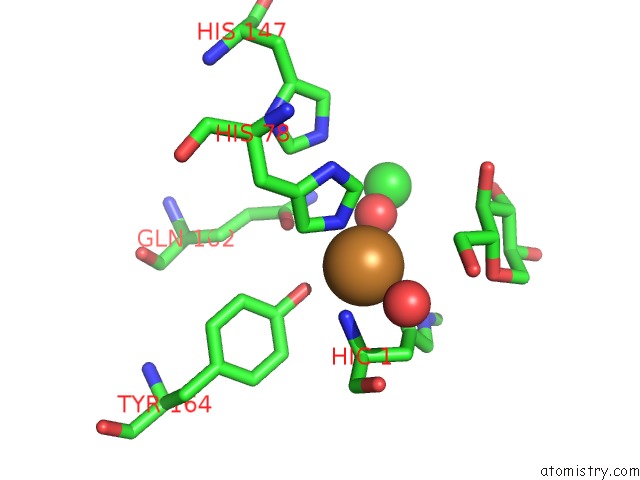
In total only one binding site of Copper was determined in the X-Ray Structure of Lpmo, PDB code: 5aci:
In total only one binding site of Copper was determined in the X-Ray Structure of Lpmo, PDB code: 5aci:
Copper binding site 1 out of 1 in 5aci
Go back to
Copper binding site 1 out
of 1 in the X-Ray Structure of Lpmo
Mono view
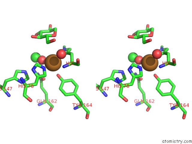
Stereo pair view
Mono view
Stereo pair view
A full contact list of Copper with other atoms in the Cu binding
site number 1 of X-Ray Structure of Lpmo within 5.0Å range:
|
Reference:
K.E.Frandsen,
T.J.Simmons,
P.Dupree,
J.C.Poulsen,
G.R.Hemsworth,
L.Ciano,
E.M.Johnston,
M.Tovborg,
K.S.Johansen,
P.Von Freiesleben,
L.Marmuse,
S.Fort,
S.Cottaz,
H.Driguez,
B.Henrissat,
N.Lenfant,
F.Tuna,
A.Baldansuren,
G.J.Davies,
L.Lo Leggio,
P.H.Walton.
The Molecular Basis of Polysaccharide Cleavage By Lytic Polysaccharide Monooxygenases. Nat. Chem. Biol. V. 12 298 2016.
ISSN: ESSN 1552-4469
PubMed: 26928935
DOI: 10.1038/NCHEMBIO.2029
Page generated: Wed Jul 31 03:48:23 2024
ISSN: ESSN 1552-4469
PubMed: 26928935
DOI: 10.1038/NCHEMBIO.2029
Last articles
Ca in 2PNCCa in 2PMY
Ca in 2PNY
Ca in 2PLY
Ca in 2PLX
Ca in 2PJP
Ca in 2PKT
Ca in 2PLT
Ca in 2PHX
Ca in 2PHW