Copper »
PDB 4tm7-4yst »
4wf5 »
Copper in PDB 4wf5: Crystal Structure of E.Coli Dsba Soaked with Compound 4
Enzymatic activity of Crystal Structure of E.Coli Dsba Soaked with Compound 4
All present enzymatic activity of Crystal Structure of E.Coli Dsba Soaked with Compound 4:
1.8.4.2;
1.8.4.2;
Protein crystallography data
The structure of Crystal Structure of E.Coli Dsba Soaked with Compound 4, PDB code: 4wf5
was solved by
L.A.Adams,
P.Sharma,
B.Mohanty,
O.V.Ilyichova,
M.D.Mulcair,
M.L.Williams,
E.C.Gleeson,
M.Totsika,
B.C.Doak,
S.Caria,
K.Rimmer,
S.R.Shouldice,
M.Vazirani,
S.J.Headey,
B.R.Plumb,
J.L.Martin,
B.Heras,
J.S.Simpson,
M.J.Scanlon,
with X-Ray Crystallography technique. A brief refinement statistics is given in the table below:
| Resolution Low / High (Å) | 34.46 / 1.45 |
| Space group | C 1 2 1 |
| Cell size a, b, c (Å), α, β, γ (°) | 118.246, 63.401, 74.402, 90.00, 126.19, 90.00 |
| R / Rfree (%) | 16 / 19.6 |
Other elements in 4wf5:
The structure of Crystal Structure of E.Coli Dsba Soaked with Compound 4 also contains other interesting chemical elements:
| Fluorine | (F) | 6 atoms |
Copper Binding Sites:
The binding sites of Copper atom in the Crystal Structure of E.Coli Dsba Soaked with Compound 4
(pdb code 4wf5). This binding sites where shown within
5.0 Angstroms radius around Copper atom.
In total only one binding site of Copper was determined in the Crystal Structure of E.Coli Dsba Soaked with Compound 4, PDB code: 4wf5:
In total only one binding site of Copper was determined in the Crystal Structure of E.Coli Dsba Soaked with Compound 4, PDB code: 4wf5:
Copper binding site 1 out of 1 in 4wf5
Go back to
Copper binding site 1 out
of 1 in the Crystal Structure of E.Coli Dsba Soaked with Compound 4
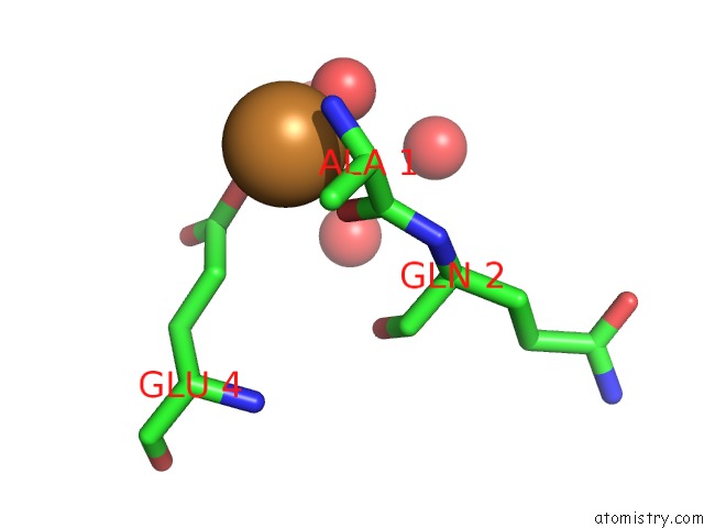
Mono view
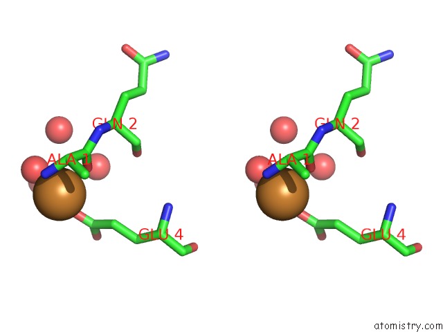
Stereo pair view
Mono view
Stereo pair view
A full contact list of Copper with other atoms in the Cu binding
site number 1 of Crystal Structure of E.Coli Dsba Soaked with Compound 4 within 5.0Å range:
|
Reference:
L.A.Adams,
P.Sharma,
B.Mohanty,
O.V.Ilyichova,
M.D.Mulcair,
M.L.Williams,
E.C.Gleeson,
M.Totsika,
B.C.Doak,
S.Caria,
K.Rimmer,
J.Horne,
S.R.Shouldice,
M.Vazirani,
S.J.Headey,
B.R.Plumb,
J.L.Martin,
B.Heras,
J.S.Simpson,
M.J.Scanlon.
Application of Fragment-Based Screening to the Design of Inhibitors of Escherichia Coli Dsba Angew.Chem.Int.Ed.Engl. 2014.
ISSN: ESSN 1521-3773
PubMed: 25556635
DOI: 10.1002/ANIE.201410341
Page generated: Mon Jul 14 04:08:35 2025
ISSN: ESSN 1521-3773
PubMed: 25556635
DOI: 10.1002/ANIE.201410341
Last articles
Fe in 1OGYFe in 1OZL
Fe in 1OZE
Fe in 1OYL
Fe in 1OYK
Fe in 1OUU
Fe in 1OVT
Fe in 1OXA
Fe in 1OVB
Fe in 1OQU