Copper »
PDB 4hhw-4mai »
4m4f »
Copper in PDB 4m4f: Radiation Damage Study of Cu T6-Insulin - 0.01 Mgy
Protein crystallography data
The structure of Radiation Damage Study of Cu T6-Insulin - 0.01 Mgy, PDB code: 4m4f
was solved by
C.G.Frankaer,
P.Harris,
K.Stahl,
with X-Ray Crystallography technique. A brief refinement statistics is given in the table below:
| Resolution Low / High (Å) | 16.28 / 1.90 |
| Space group | H 3 |
| Cell size a, b, c (Å), α, β, γ (°) | 81.130, 81.130, 33.469, 90.00, 90.00, 120.00 |
| R / Rfree (%) | 16.6 / 23.5 |
Copper Binding Sites:
The binding sites of Copper atom in the Radiation Damage Study of Cu T6-Insulin - 0.01 Mgy
(pdb code 4m4f). This binding sites where shown within
5.0 Angstroms radius around Copper atom.
In total 2 binding sites of Copper where determined in the Radiation Damage Study of Cu T6-Insulin - 0.01 Mgy, PDB code: 4m4f:
Jump to Copper binding site number: 1; 2;
In total 2 binding sites of Copper where determined in the Radiation Damage Study of Cu T6-Insulin - 0.01 Mgy, PDB code: 4m4f:
Jump to Copper binding site number: 1; 2;
Copper binding site 1 out of 2 in 4m4f
Go back to
Copper binding site 1 out
of 2 in the Radiation Damage Study of Cu T6-Insulin - 0.01 Mgy
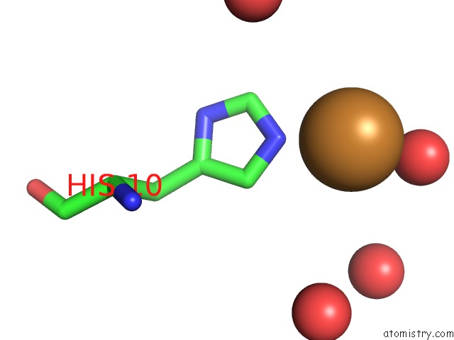
Mono view
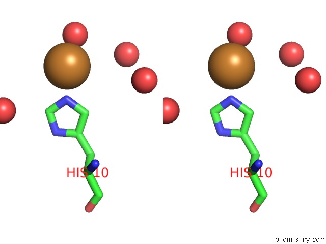
Stereo pair view
Mono view
Stereo pair view
A full contact list of Copper with other atoms in the Cu binding
site number 1 of Radiation Damage Study of Cu T6-Insulin - 0.01 Mgy within 5.0Å range:
|
Copper binding site 2 out of 2 in 4m4f
Go back to
Copper binding site 2 out
of 2 in the Radiation Damage Study of Cu T6-Insulin - 0.01 Mgy
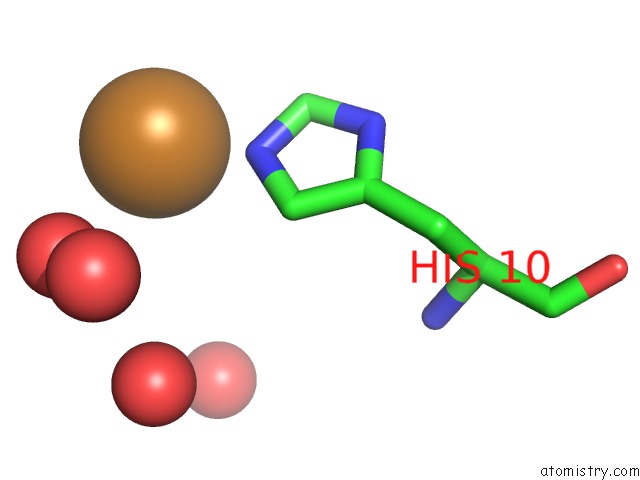
Mono view
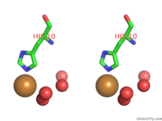
Stereo pair view
Mono view
Stereo pair view
A full contact list of Copper with other atoms in the Cu binding
site number 2 of Radiation Damage Study of Cu T6-Insulin - 0.01 Mgy within 5.0Å range:
|
Reference:
C.G.Frankaer,
S.Mossin,
K.Stahl,
P.Harris.
Towards Accurate Structural Characterization of Metal Centres in Protein Crystals: the Structures of Ni and Cu T6 Bovine Insulin Derivatives. Acta Crystallogr.,Sect.D V. 70 110 2014.
ISSN: ISSN 0907-4449
PubMed: 24419384
DOI: 10.1107/S1399004713029040
Page generated: Wed Jul 31 03:14:05 2024
ISSN: ISSN 0907-4449
PubMed: 24419384
DOI: 10.1107/S1399004713029040
Last articles
Zn in 9JYWZn in 9IR4
Zn in 9IR3
Zn in 9GMX
Zn in 9GMW
Zn in 9JEJ
Zn in 9ERF
Zn in 9ERE
Zn in 9EGV
Zn in 9EGW