Copper »
PDB 4hhw-4mai »
4hip »
Copper in PDB 4hip: Crystal Structure of the Pseudomonas Aeruginosa Azurin, H126NO YOH109
Protein crystallography data
The structure of Crystal Structure of the Pseudomonas Aeruginosa Azurin, H126NO YOH109, PDB code: 4hip
was solved by
N.Herrera,
J.J.Warren,
H.B.Gray,
with X-Ray Crystallography technique. A brief refinement statistics is given in the table below:
| Resolution Low / High (Å) | 36.50 / 1.90 |
| Space group | P 2 21 21 |
| Cell size a, b, c (Å), α, β, γ (°) | 49.546, 65.804, 73.005, 90.00, 90.00, 90.00 |
| R / Rfree (%) | 25 / 31.7 |
Copper Binding Sites:
The binding sites of Copper atom in the Crystal Structure of the Pseudomonas Aeruginosa Azurin, H126NO YOH109
(pdb code 4hip). This binding sites where shown within
5.0 Angstroms radius around Copper atom.
In total 2 binding sites of Copper where determined in the Crystal Structure of the Pseudomonas Aeruginosa Azurin, H126NO YOH109, PDB code: 4hip:
Jump to Copper binding site number: 1; 2;
In total 2 binding sites of Copper where determined in the Crystal Structure of the Pseudomonas Aeruginosa Azurin, H126NO YOH109, PDB code: 4hip:
Jump to Copper binding site number: 1; 2;
Copper binding site 1 out of 2 in 4hip
Go back to
Copper binding site 1 out
of 2 in the Crystal Structure of the Pseudomonas Aeruginosa Azurin, H126NO YOH109
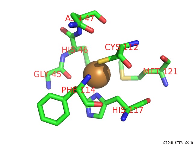
Mono view
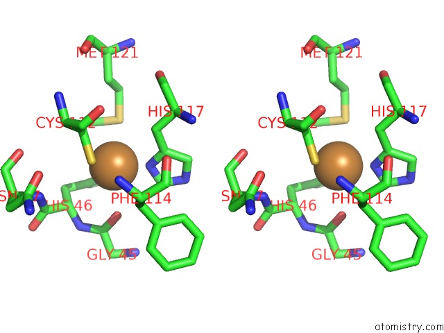
Stereo pair view
Mono view
Stereo pair view
A full contact list of Copper with other atoms in the Cu binding
site number 1 of Crystal Structure of the Pseudomonas Aeruginosa Azurin, H126NO YOH109 within 5.0Å range:
|
Copper binding site 2 out of 2 in 4hip
Go back to
Copper binding site 2 out
of 2 in the Crystal Structure of the Pseudomonas Aeruginosa Azurin, H126NO YOH109
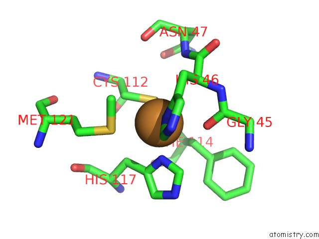
Mono view
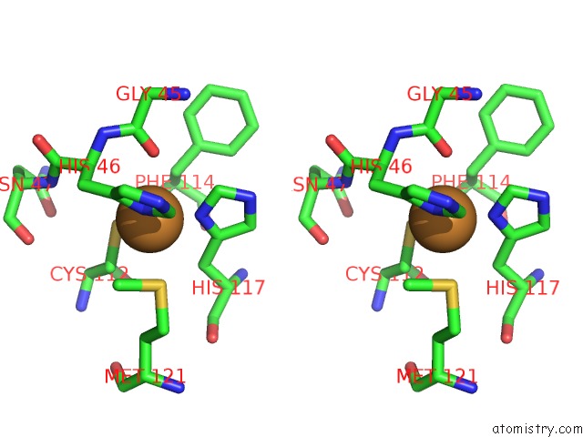
Stereo pair view
Mono view
Stereo pair view
A full contact list of Copper with other atoms in the Cu binding
site number 2 of Crystal Structure of the Pseudomonas Aeruginosa Azurin, H126NO YOH109 within 5.0Å range:
|
Reference:
J.J.Warren,
N.Herrera,
M.G.Hill,
J.R.Winkler,
H.B.Gray.
Electron Flow Through Nitrotyrosinate in Pseudomonas Aeruginosa Azurin. J.Am.Chem.Soc. V. 135 11151 2013.
ISSN: ISSN 0002-7863
PubMed: 23859602
DOI: 10.1021/JA403734N
Page generated: Mon Jul 14 03:46:08 2025
ISSN: ISSN 0002-7863
PubMed: 23859602
DOI: 10.1021/JA403734N
Last articles
F in 7L5EF in 7L72
F in 7L5P
F in 7L69
F in 7L5O
F in 7L0K
F in 7L4W
F in 7L4U
F in 7L4N
F in 7L4M