Copper »
PDB 4b61-4e9t »
4dz0 »
Copper in PDB 4dz0: Crystal Structure of the Cu-Adduct of Human H-Ferritin Variant MIC1 Labeled with A Dansyl Fluorophore
Enzymatic activity of Crystal Structure of the Cu-Adduct of Human H-Ferritin Variant MIC1 Labeled with A Dansyl Fluorophore
All present enzymatic activity of Crystal Structure of the Cu-Adduct of Human H-Ferritin Variant MIC1 Labeled with A Dansyl Fluorophore:
1.16.3.1;
1.16.3.1;
Protein crystallography data
The structure of Crystal Structure of the Cu-Adduct of Human H-Ferritin Variant MIC1 Labeled with A Dansyl Fluorophore, PDB code: 4dz0
was solved by
F.A.Tezcan,
D.J.E.Huard,
with X-Ray Crystallography technique. A brief refinement statistics is given in the table below:
| Resolution Low / High (Å) | 36.84 / 2.50 |
| Space group | F 4 3 2 |
| Cell size a, b, c (Å), α, β, γ (°) | 180.434, 180.434, 180.434, 90.00, 90.00, 90.00 |
| R / Rfree (%) | 22.4 / 30 |
Other elements in 4dz0:
The structure of Crystal Structure of the Cu-Adduct of Human H-Ferritin Variant MIC1 Labeled with A Dansyl Fluorophore also contains other interesting chemical elements:
| Calcium | (Ca) | 3 atoms |
Copper Binding Sites:
The binding sites of Copper atom in the Crystal Structure of the Cu-Adduct of Human H-Ferritin Variant MIC1 Labeled with A Dansyl Fluorophore
(pdb code 4dz0). This binding sites where shown within
5.0 Angstroms radius around Copper atom.
In total 3 binding sites of Copper where determined in the Crystal Structure of the Cu-Adduct of Human H-Ferritin Variant MIC1 Labeled with A Dansyl Fluorophore, PDB code: 4dz0:
Jump to Copper binding site number: 1; 2; 3;
In total 3 binding sites of Copper where determined in the Crystal Structure of the Cu-Adduct of Human H-Ferritin Variant MIC1 Labeled with A Dansyl Fluorophore, PDB code: 4dz0:
Jump to Copper binding site number: 1; 2; 3;
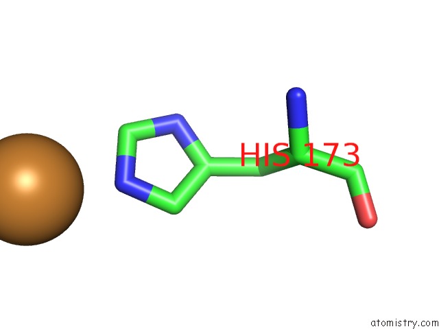
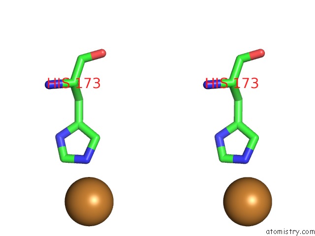
Copper binding site 1 out of 3 in 4dz0
Go back to
Copper binding site 1 out
of 3 in the Crystal Structure of the Cu-Adduct of Human H-Ferritin Variant MIC1 Labeled with A Dansyl Fluorophore
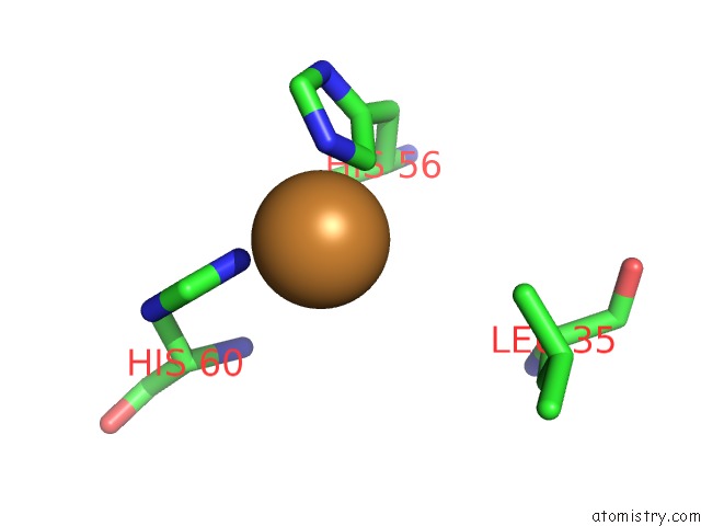
Mono view
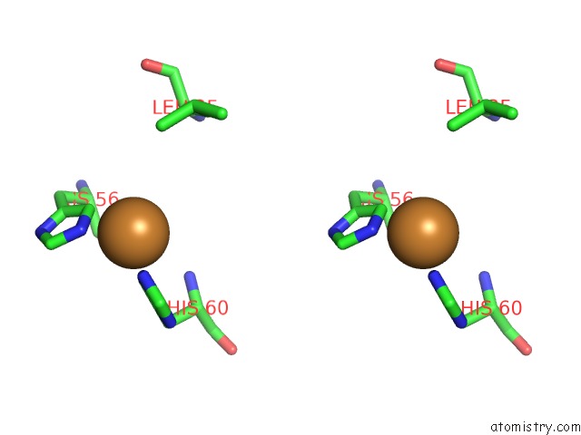
Stereo pair view
Mono view
Stereo pair view
A full contact list of Copper with other atoms in the Cu binding
site number 1 of Crystal Structure of the Cu-Adduct of Human H-Ferritin Variant MIC1 Labeled with A Dansyl Fluorophore within 5.0Å range:
|
Copper binding site 2 out of 3 in 4dz0
Go back to
Copper binding site 2 out
of 3 in the Crystal Structure of the Cu-Adduct of Human H-Ferritin Variant MIC1 Labeled with A Dansyl Fluorophore
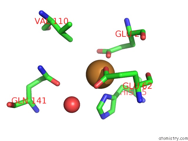
Mono view
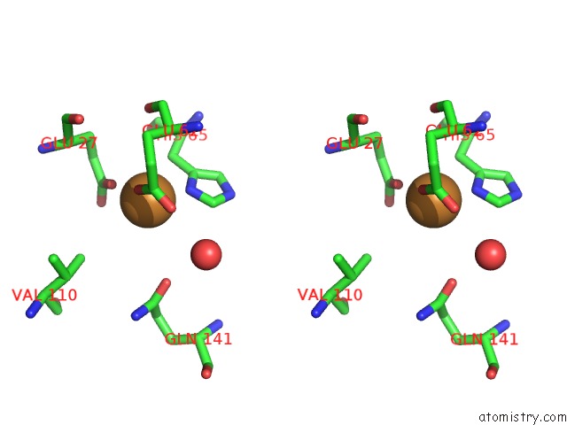
Stereo pair view
Mono view
Stereo pair view
A full contact list of Copper with other atoms in the Cu binding
site number 2 of Crystal Structure of the Cu-Adduct of Human H-Ferritin Variant MIC1 Labeled with A Dansyl Fluorophore within 5.0Å range:
|
Copper binding site 3 out of 3 in 4dz0
Go back to
Copper binding site 3 out
of 3 in the Crystal Structure of the Cu-Adduct of Human H-Ferritin Variant MIC1 Labeled with A Dansyl Fluorophore
Mono view
Stereo pair view
Mono view
Stereo pair view
A full contact list of Copper with other atoms in the Cu binding
site number 3 of Crystal Structure of the Cu-Adduct of Human H-Ferritin Variant MIC1 Labeled with A Dansyl Fluorophore within 5.0Å range:
|
Reference:
D.J.Huard,
K.M.Kane,
F.A.Tezcan.
Re-Engineering Protein Interfaces Yields Copper-Inducible Ferritin Cage Assembly. Nat.Chem.Biol. V. 9 169 2013.
ISSN: ISSN 1552-4450
PubMed: 23340339
DOI: 10.1038/NCHEMBIO.1163
Page generated: Mon Jul 14 03:31:12 2025
ISSN: ISSN 1552-4450
PubMed: 23340339
DOI: 10.1038/NCHEMBIO.1163
Last articles
Fe in 2YXOFe in 2YRS
Fe in 2YXC
Fe in 2YNM
Fe in 2YVJ
Fe in 2YP1
Fe in 2YU2
Fe in 2YU1
Fe in 2YQB
Fe in 2YOO