Copper »
PDB 3mnd-3qjo »
3paz »
Copper in PDB 3paz: Reduced Native Pseudoazurin From A. Faecalis
Protein crystallography data
The structure of Reduced Native Pseudoazurin From A. Faecalis, PDB code: 3paz
was solved by
E.T.Adman,
C.A.P.Libeu,
with X-Ray Crystallography technique. A brief refinement statistics is given in the table below:
| Resolution Low / High (Å) | 22.30 / 1.73 |
| Space group | P 65 |
| Cell size a, b, c (Å), α, β, γ (°) | 50.060, 50.060, 98.720, 90.00, 90.00, 120.00 |
| R / Rfree (%) | 16.4 / n/a |
Copper Binding Sites:
The binding sites of Copper atom in the Reduced Native Pseudoazurin From A. Faecalis
(pdb code 3paz). This binding sites where shown within
5.0 Angstroms radius around Copper atom.
In total only one binding site of Copper was determined in the Reduced Native Pseudoazurin From A. Faecalis, PDB code: 3paz:
In total only one binding site of Copper was determined in the Reduced Native Pseudoazurin From A. Faecalis, PDB code: 3paz:
Copper binding site 1 out of 1 in 3paz
Go back to
Copper binding site 1 out
of 1 in the Reduced Native Pseudoazurin From A. Faecalis
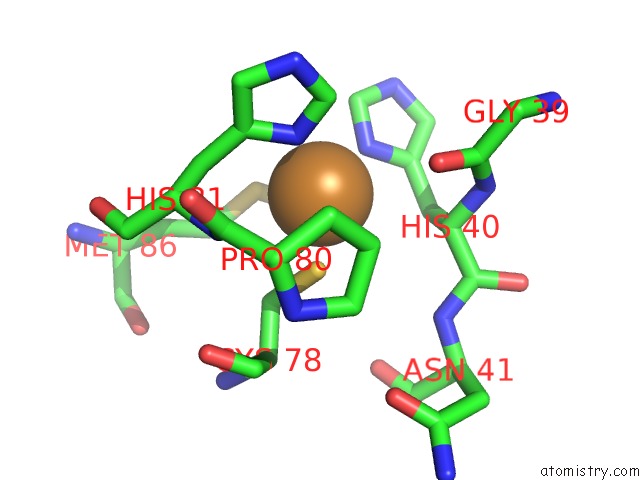
Mono view
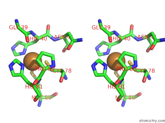
Stereo pair view
Mono view
Stereo pair view
A full contact list of Copper with other atoms in the Cu binding
site number 1 of Reduced Native Pseudoazurin From A. Faecalis within 5.0Å range:
|
Reference:
C.A.Libeu,
M.Kukimoto,
M.Nishiyama,
S.Horinouchi,
E.T.Adman.
Site-Directed Mutants of Pseudoazurin: Explanation of Increased Redox Potentials From X-Ray Structures and From Calculation of Redox Potential Differences. Biochemistry V. 36 13160 1997.
ISSN: ISSN 0006-2960
PubMed: 9341204
DOI: 10.1021/BI9704111
Page generated: Wed Jul 31 01:31:40 2024
ISSN: ISSN 0006-2960
PubMed: 9341204
DOI: 10.1021/BI9704111
Last articles
Zn in 9MJ5Zn in 9HNW
Zn in 9G0L
Zn in 9FNE
Zn in 9DZN
Zn in 9E0I
Zn in 9D32
Zn in 9DAK
Zn in 8ZXC
Zn in 8ZUF