Copper »
PDB 3iea-3mn0 »
3in2 »
Copper in PDB 3in2: Crystal Structure of the N47S/M121L Variant of Pseudomonas Aeruginosa Azurin in the Cu(II) State
Protein crystallography data
The structure of Crystal Structure of the N47S/M121L Variant of Pseudomonas Aeruginosa Azurin in the Cu(II) State, PDB code: 3in2
was solved by
Y.G.Gao,
H.Robinson,
with X-Ray Crystallography technique. A brief refinement statistics is given in the table below:
| Resolution Low / High (Å) | 10.00 / 2.60 |
| Space group | P 43 21 2 |
| Cell size a, b, c (Å), α, β, γ (°) | 49.660, 49.660, 105.100, 90.00, 90.00, 90.00 |
| R / Rfree (%) | 23.8 / 26.6 |
Copper Binding Sites:
The binding sites of Copper atom in the Crystal Structure of the N47S/M121L Variant of Pseudomonas Aeruginosa Azurin in the Cu(II) State
(pdb code 3in2). This binding sites where shown within
5.0 Angstroms radius around Copper atom.
In total only one binding site of Copper was determined in the Crystal Structure of the N47S/M121L Variant of Pseudomonas Aeruginosa Azurin in the Cu(II) State, PDB code: 3in2:
In total only one binding site of Copper was determined in the Crystal Structure of the N47S/M121L Variant of Pseudomonas Aeruginosa Azurin in the Cu(II) State, PDB code: 3in2:
Copper binding site 1 out of 1 in 3in2
Go back to
Copper binding site 1 out
of 1 in the Crystal Structure of the N47S/M121L Variant of Pseudomonas Aeruginosa Azurin in the Cu(II) State
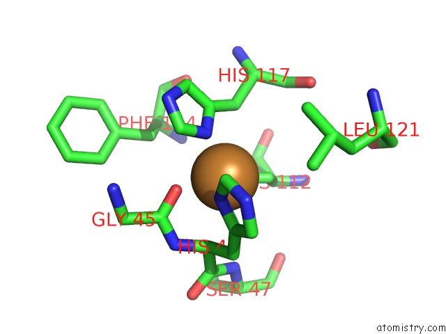
Mono view
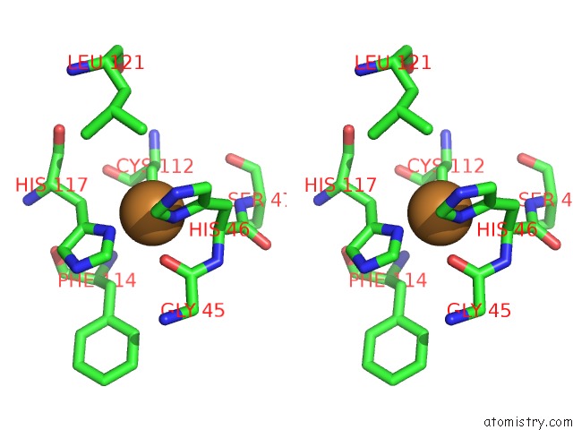
Stereo pair view
Mono view
Stereo pair view
A full contact list of Copper with other atoms in the Cu binding
site number 1 of Crystal Structure of the N47S/M121L Variant of Pseudomonas Aeruginosa Azurin in the Cu(II) State within 5.0Å range:
|
Reference:
N.M.Marshall,
D.K.Garner,
T.D.Wilson,
Y.G.Gao,
H.Robinson,
M.J.Nilges,
Y.Lu.
Rationally Tuning the Reduction Potential of A Single Cupredoxin Beyond the Natural Range. Nature V. 462 113 2009.
ISSN: ISSN 0028-0836
PubMed: 19890331
DOI: 10.1038/NATURE08551
Page generated: Wed Jul 31 01:12:59 2024
ISSN: ISSN 0028-0836
PubMed: 19890331
DOI: 10.1038/NATURE08551
Last articles
Ca in 5SWICa in 5SVE
Ca in 5SSX
Ca in 5SV0
Ca in 5STD
Ca in 5SSZ
Ca in 5SSY
Ca in 5SIC
Ca in 5SBD
Ca in 5SBE