Copper »
PDB 3f00-3ie9 »
3hnl »
Copper in PDB 3hnl: Crystal Structure of the Cu-Induced Dimer of the Engineered Cyt CB562 Variant Ridc-1
Protein crystallography data
The structure of Crystal Structure of the Cu-Induced Dimer of the Engineered Cyt CB562 Variant Ridc-1, PDB code: 3hnl
was solved by
E.N.Salgado,
R.A.Lewis,
J.Brodin,
F.A.Tezcan,
with X-Ray Crystallography technique. A brief refinement statistics is given in the table below:
| Resolution Low / High (Å) | 22.20 / 2.20 |
| Space group | C 2 2 21 |
| Cell size a, b, c (Å), α, β, γ (°) | 66.165, 87.045, 80.765, 90.00, 90.00, 90.00 |
| R / Rfree (%) | 20.4 / 27 |
Other elements in 3hnl:
The structure of Crystal Structure of the Cu-Induced Dimer of the Engineered Cyt CB562 Variant Ridc-1 also contains other interesting chemical elements:
| Iron | (Fe) | 2 atoms |
| Chlorine | (Cl) | 2 atoms |
Copper Binding Sites:
The binding sites of Copper atom in the Crystal Structure of the Cu-Induced Dimer of the Engineered Cyt CB562 Variant Ridc-1
(pdb code 3hnl). This binding sites where shown within
5.0 Angstroms radius around Copper atom.
In total 2 binding sites of Copper where determined in the Crystal Structure of the Cu-Induced Dimer of the Engineered Cyt CB562 Variant Ridc-1, PDB code: 3hnl:
Jump to Copper binding site number: 1; 2;
In total 2 binding sites of Copper where determined in the Crystal Structure of the Cu-Induced Dimer of the Engineered Cyt CB562 Variant Ridc-1, PDB code: 3hnl:
Jump to Copper binding site number: 1; 2;
Copper binding site 1 out of 2 in 3hnl
Go back to
Copper binding site 1 out
of 2 in the Crystal Structure of the Cu-Induced Dimer of the Engineered Cyt CB562 Variant Ridc-1
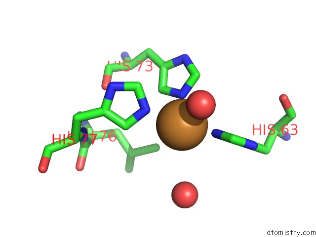
Mono view

Stereo pair view
Mono view
Stereo pair view
A full contact list of Copper with other atoms in the Cu binding
site number 1 of Crystal Structure of the Cu-Induced Dimer of the Engineered Cyt CB562 Variant Ridc-1 within 5.0Å range:
|
Copper binding site 2 out of 2 in 3hnl
Go back to
Copper binding site 2 out
of 2 in the Crystal Structure of the Cu-Induced Dimer of the Engineered Cyt CB562 Variant Ridc-1

Mono view

Stereo pair view
Mono view
Stereo pair view
A full contact list of Copper with other atoms in the Cu binding
site number 2 of Crystal Structure of the Cu-Induced Dimer of the Engineered Cyt CB562 Variant Ridc-1 within 5.0Å range:
|
Reference:
E.N.Salgado,
X.I.Ambroggio,
J.D.Brodin,
R.A.Lewis,
B.Kuhlman,
F.A.Tezcan.
Metal Templated Design of Protein Interfaces. Proc.Natl.Acad.Sci.Usa V. 107 1827 2010.
ISSN: ISSN 0027-8424
PubMed: 20080561
DOI: 10.1073/PNAS.0906852107
Page generated: Wed Jul 31 01:04:05 2024
ISSN: ISSN 0027-8424
PubMed: 20080561
DOI: 10.1073/PNAS.0906852107
Last articles
Zn in 9MJ5Zn in 9HNW
Zn in 9G0L
Zn in 9FNE
Zn in 9DZN
Zn in 9E0I
Zn in 9D32
Zn in 9DAK
Zn in 8ZXC
Zn in 8ZUF