Copper »
PDB 3f00-3ie9 »
3fq1 »
Copper in PDB 3fq1: Azurin C112D/M121I
Protein crystallography data
The structure of Azurin C112D/M121I, PDB code: 3fq1
was solved by
K.M.Lancaster,
H.B.Gray,
with X-Ray Crystallography technique. A brief refinement statistics is given in the table below:
| Resolution Low / High (Å) | 19.99 / 1.90 |
| Space group | C 2 2 21 |
| Cell size a, b, c (Å), α, β, γ (°) | 48.500, 54.800, 95.780, 90.00, 90.00, 90.00 |
| R / Rfree (%) | 19.8 / 25.6 |
Copper Binding Sites:
The binding sites of Copper atom in the Azurin C112D/M121I
(pdb code 3fq1). This binding sites where shown within
5.0 Angstroms radius around Copper atom.
In total 2 binding sites of Copper where determined in the Azurin C112D/M121I, PDB code: 3fq1:
Jump to Copper binding site number: 1; 2;
In total 2 binding sites of Copper where determined in the Azurin C112D/M121I, PDB code: 3fq1:
Jump to Copper binding site number: 1; 2;
Copper binding site 1 out of 2 in 3fq1
Go back to
Copper binding site 1 out
of 2 in the Azurin C112D/M121I
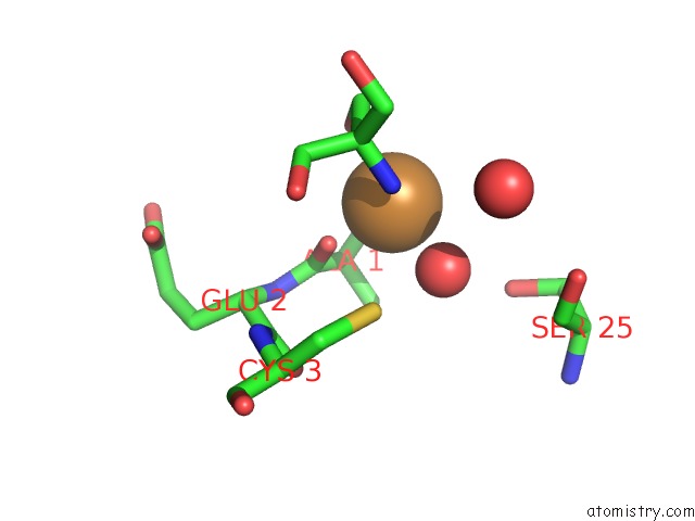
Mono view
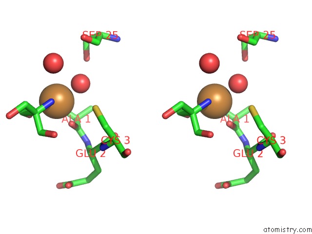
Stereo pair view
Mono view
Stereo pair view
A full contact list of Copper with other atoms in the Cu binding
site number 1 of Azurin C112D/M121I within 5.0Å range:
|
Copper binding site 2 out of 2 in 3fq1
Go back to
Copper binding site 2 out
of 2 in the Azurin C112D/M121I
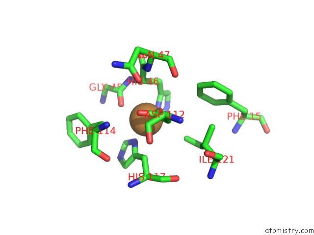
Mono view
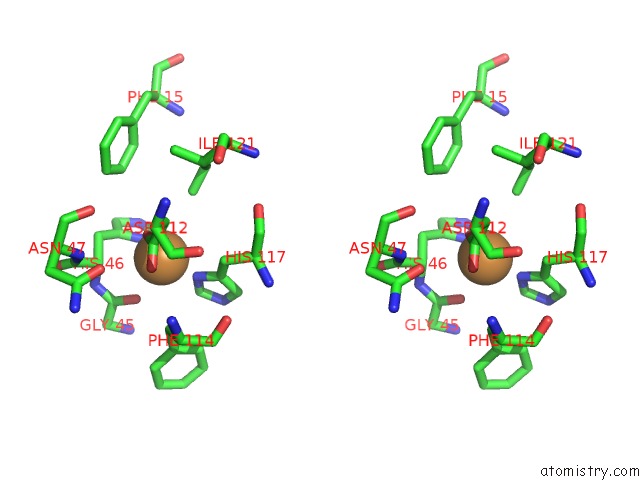
Stereo pair view
Mono view
Stereo pair view
A full contact list of Copper with other atoms in the Cu binding
site number 2 of Azurin C112D/M121I within 5.0Å range:
|
Reference:
K.M.Lancaster,
S.Debeer George,
K.Yokoyama,
J.H.Richards,
H.B.Gray.
Type Zero Copper Proteins. Nat Chem V. 1 711 2009.
ISSN: ISSN 1755-4349
PubMed: 20305734
DOI: 10.1038/NCHEM.412
Page generated: Wed Jul 31 00:54:28 2024
ISSN: ISSN 1755-4349
PubMed: 20305734
DOI: 10.1038/NCHEM.412
Last articles
Zn in 9MJ5Zn in 9HNW
Zn in 9G0L
Zn in 9FNE
Zn in 9DZN
Zn in 9E0I
Zn in 9D32
Zn in 9DAK
Zn in 8ZXC
Zn in 8ZUF