Copper in PDB 3cqp: Human SOD1 G85R Variant, Structure I
Enzymatic activity of Human SOD1 G85R Variant, Structure I
All present enzymatic activity of Human SOD1 G85R Variant, Structure I:
1.15.1.1;
Protein crystallography data
The structure of Human SOD1 G85R Variant, Structure I, PDB code: 3cqp
was solved by
X.Cao,
S.Antonyuk,
S.V.Seetharaman,
L.J.Whitson,
A.B.Taylor,
S.P.Holloway,
R.W.Strange,
P.A.Doucette,
J.S.Valentine,
A.Tiwari,
L.J.Hayward,
S.Padua,
J.A.Cohlberg,
S.S.Hasnain,
P.J.Hart,
with X-Ray Crystallography technique. A brief refinement statistics is given in the table below:
Resolution Low / High (Å)
|
50.00 /
1.95
|
Space group
|
I 21 21 21
|
Cell size a, b, c (Å), α, β, γ (°)
|
73.472,
116.808,
147.764,
90.00,
90.00,
90.00
|
R / Rfree (%)
|
18.3 /
22.3
|
Other elements in 3cqp:
The structure of Human SOD1 G85R Variant, Structure I also contains other interesting chemical elements:
Copper Binding Sites:
The binding sites of Copper atom in the Human SOD1 G85R Variant, Structure I
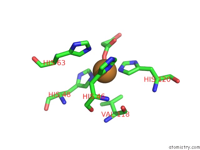
(pdb code 3cqp). This binding sites where shown within
5.0 Angstroms radius around Copper atom.
In total 4 binding sites of Copper where determined in the
Human SOD1 G85R Variant, Structure I, PDB code: 3cqp:
Jump to Copper binding site number:
1;
2;
3;
4;
Copper binding site 1 out
of 4 in 3cqp
Go back to
Copper Binding Sites List in 3cqp
Copper binding site 1 out
of 4 in the Human SOD1 G85R Variant, Structure I
 Mono view
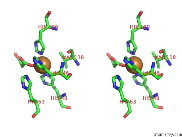
 Stereo pair view
|
A full contact list of Copper with other atoms in the Cu binding
site number 1 of Human SOD1 G85R Variant, Structure I within 5.0Å range:
probe
|
atom
|
residue
|
distance (Å)
|
B
|
Occ
|
A:Cu154
b:46.5
occ:0.80
|
O6
|
A:MLI157
|
1.8
|
49.6
|
1.0
|
NE2
|
A:HIS120
|
1.9
|
16.0
|
1.0
|
NE2
|
A:HIS48
|
2.0
|
17.2
|
1.0
|
ND1
|
A:HIS46
|
2.4
|
19.9
|
1.0
|
CD2
|
A:HIS120
|
2.8
|
16.2
|
1.0
|
CE1
|
A:HIS48
|
2.9
|
17.8
|
1.0
|
C2
|
A:MLI157
|
2.9
|
49.4
|
1.0
|
CE1
|
A:HIS120
|
3.0
|
15.9
|
1.0
|
CD2
|
A:HIS48
|
3.1
|
17.1
|
1.0
|
CG
|
A:HIS46
|
3.2
|
17.9
|
1.0
|
NE2
|
A:HIS63
|
3.3
|
18.9
|
1.0
|
CE1
|
A:HIS46
|
3.4
|
19.6
|
1.0
|
CB
|
A:HIS46
|
3.4
|
16.8
|
1.0
|
CD2
|
A:HIS63
|
3.5
|
18.9
|
1.0
|
O7
|
A:MLI157
|
3.7
|
48.8
|
1.0
|
C1
|
A:MLI157
|
3.8
|
49.5
|
1.0
|
CG
|
A:HIS120
|
4.0
|
16.6
|
1.0
|
ND1
|
A:HIS120
|
4.0
|
17.6
|
1.0
|
ND1
|
A:HIS48
|
4.1
|
18.2
|
1.0
|
CB
|
A:VAL118
|
4.1
|
15.1
|
1.0
|
CG
|
A:HIS48
|
4.2
|
17.3
|
1.0
|
CG1
|
A:VAL118
|
4.3
|
15.4
|
1.0
|
CD2
|
A:HIS46
|
4.4
|
17.9
|
1.0
|
CE1
|
A:HIS63
|
4.4
|
18.5
|
1.0
|
NE2
|
A:HIS46
|
4.4
|
18.7
|
1.0
|
CA
|
A:HIS46
|
4.5
|
16.1
|
1.0
|
N
|
A:HIS46
|
4.6
|
15.7
|
1.0
|
CG
|
A:HIS63
|
4.7
|
17.1
|
1.0
|
O
|
A:VAL118
|
4.8
|
15.3
|
1.0
|
CG2
|
A:VAL118
|
4.8
|
14.2
|
1.0
|
C3
|
A:MLI157
|
4.8
|
51.3
|
1.0
|
O
|
A:HIS46
|
4.9
|
15.8
|
1.0
|
C
|
A:HIS46
|
5.0
|
15.7
|
1.0
|
|
Copper binding site 2 out
of 4 in 3cqp
Go back to
Copper Binding Sites List in 3cqp
Copper binding site 2 out
of 4 in the Human SOD1 G85R Variant, Structure I
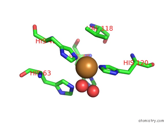
 Mono view
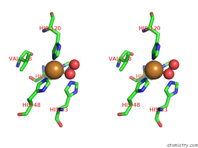
 Stereo pair view
|
A full contact list of Copper with other atoms in the Cu binding
site number 2 of Human SOD1 G85R Variant, Structure I within 5.0Å range:
probe
|
atom
|
residue
|
distance (Å)
|
B
|
Occ
|
B:Cu154
b:43.3
occ:0.60
|
NE2
|
B:HIS120
|
1.9
|
20.1
|
1.0
|
NE2
|
B:HIS48
|
2.0
|
16.8
|
1.0
|
ND1
|
B:HIS46
|
2.3
|
18.3
|
1.0
|
CD2
|
B:HIS120
|
2.8
|
17.5
|
1.0
|
CE1
|
B:HIS48
|
2.9
|
15.7
|
1.0
|
CE1
|
B:HIS120
|
2.9
|
19.5
|
1.0
|
NE2
|
B:HIS63
|
3.1
|
17.4
|
1.0
|
CG
|
B:HIS46
|
3.1
|
16.9
|
1.0
|
CD2
|
B:HIS48
|
3.1
|
14.4
|
1.0
|
O
|
B:HOH177
|
3.2
|
33.0
|
1.0
|
CB
|
B:HIS46
|
3.2
|
16.6
|
1.0
|
CE1
|
B:HIS46
|
3.3
|
20.2
|
1.0
|
CD2
|
B:HIS63
|
3.4
|
17.9
|
1.0
|
O
|
B:HOH232
|
3.9
|
42.7
|
1.0
|
CG
|
B:HIS120
|
3.9
|
16.8
|
1.0
|
ND1
|
B:HIS120
|
4.0
|
19.1
|
1.0
|
ND1
|
B:HIS48
|
4.0
|
17.0
|
1.0
|
CE1
|
B:HIS63
|
4.1
|
17.8
|
1.0
|
CG
|
B:HIS48
|
4.2
|
15.3
|
1.0
|
CB
|
B:VAL118
|
4.2
|
14.8
|
1.0
|
CD2
|
B:HIS46
|
4.3
|
17.4
|
1.0
|
CA
|
B:HIS46
|
4.3
|
16.3
|
1.0
|
NE2
|
B:HIS46
|
4.3
|
17.4
|
1.0
|
N
|
B:HIS46
|
4.4
|
16.2
|
1.0
|
CG1
|
B:VAL118
|
4.5
|
16.2
|
1.0
|
CG
|
B:HIS63
|
4.6
|
17.6
|
1.0
|
O
|
B:HIS46
|
4.8
|
15.7
|
1.0
|
O
|
B:VAL118
|
4.8
|
14.8
|
1.0
|
C
|
B:HIS46
|
4.8
|
15.9
|
1.0
|
ND1
|
B:HIS63
|
4.9
|
17.4
|
1.0
|
CG2
|
B:VAL118
|
4.9
|
14.5
|
1.0
|
|
Copper binding site 3 out
of 4 in 3cqp
Go back to
Copper Binding Sites List in 3cqp
Copper binding site 3 out
of 4 in the Human SOD1 G85R Variant, Structure I
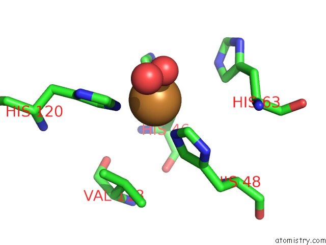
 Mono view
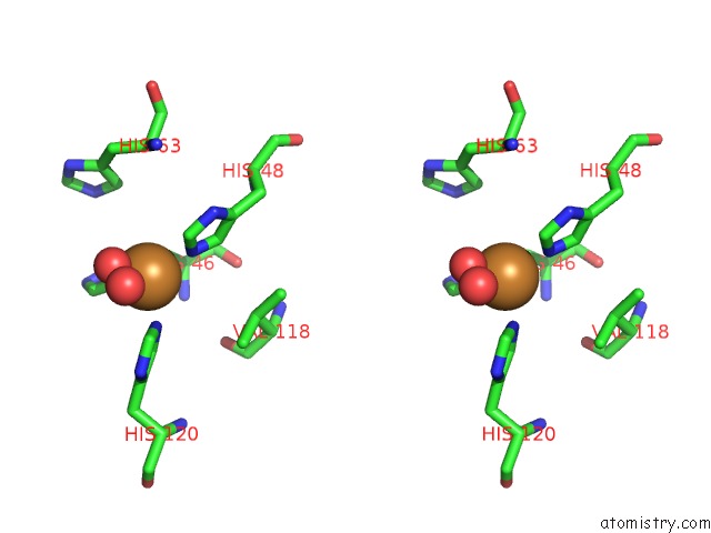
 Stereo pair view
|
A full contact list of Copper with other atoms in the Cu binding
site number 3 of Human SOD1 G85R Variant, Structure I within 5.0Å range:
probe
|
atom
|
residue
|
distance (Å)
|
B
|
Occ
|
C:Cu154
b:32.8
occ:1.00
|
NE2
|
C:HIS120
|
2.1
|
24.7
|
1.0
|
NE2
|
C:HIS48
|
2.1
|
24.5
|
1.0
|
ND1
|
C:HIS46
|
2.3
|
25.8
|
1.0
|
O
|
C:HOH204
|
2.4
|
34.8
|
1.0
|
CE1
|
C:HIS48
|
2.9
|
24.2
|
1.0
|
CE1
|
C:HIS120
|
3.0
|
24.9
|
1.0
|
CD2
|
C:HIS120
|
3.1
|
24.7
|
1.0
|
CG
|
C:HIS46
|
3.3
|
25.3
|
1.0
|
CE1
|
C:HIS46
|
3.3
|
28.4
|
1.0
|
CD2
|
C:HIS48
|
3.3
|
22.5
|
1.0
|
CB
|
C:HIS46
|
3.5
|
23.5
|
1.0
|
ND1
|
C:HIS48
|
4.1
|
23.9
|
1.0
|
ND1
|
C:HIS120
|
4.1
|
24.7
|
1.0
|
CG
|
C:HIS120
|
4.1
|
25.1
|
1.0
|
CG
|
C:HIS48
|
4.3
|
22.2
|
1.0
|
O
|
C:HOH177
|
4.4
|
43.8
|
1.0
|
NE2
|
C:HIS46
|
4.4
|
27.9
|
1.0
|
CD2
|
C:HIS46
|
4.4
|
27.0
|
1.0
|
CG1
|
C:VAL118
|
4.7
|
21.0
|
1.0
|
CD2
|
C:HIS63
|
4.8
|
33.3
|
1.0
|
CB
|
C:VAL118
|
4.8
|
20.1
|
1.0
|
CA
|
C:HIS46
|
4.9
|
22.8
|
1.0
|
|
Copper binding site 4 out
of 4 in 3cqp
Go back to
Copper Binding Sites List in 3cqp
Copper binding site 4 out
of 4 in the Human SOD1 G85R Variant, Structure I
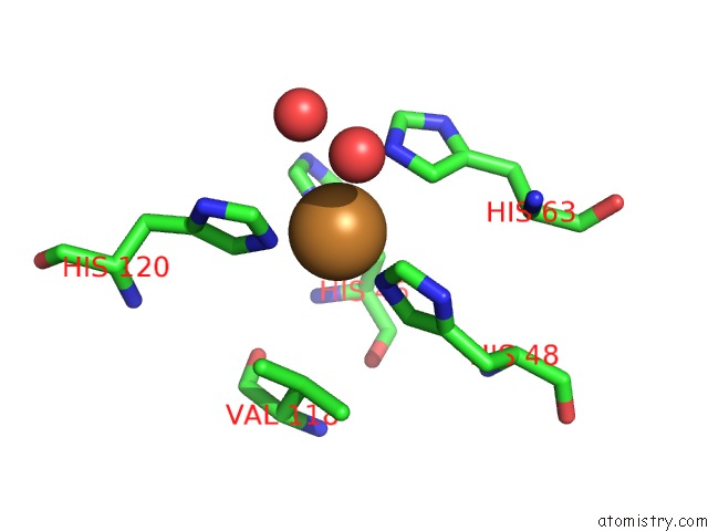
 Mono view
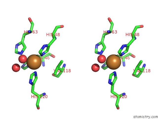
 Stereo pair view
|
A full contact list of Copper with other atoms in the Cu binding
site number 4 of Human SOD1 G85R Variant, Structure I within 5.0Å range:
probe
|
atom
|
residue
|
distance (Å)
|
B
|
Occ
|
D:Cu154
b:47.5
occ:0.80
|
NE2
|
D:HIS48
|
2.0
|
19.9
|
1.0
|
NE2
|
D:HIS120
|
2.0
|
21.0
|
1.0
|
ND1
|
D:HIS46
|
2.3
|
21.9
|
1.0
|
CE1
|
D:HIS48
|
2.7
|
19.3
|
1.0
|
NE2
|
D:HIS63
|
2.9
|
24.6
|
1.0
|
CE1
|
D:HIS120
|
2.9
|
21.4
|
1.0
|
CD2
|
D:HIS120
|
3.0
|
20.1
|
1.0
|
CD2
|
D:HIS48
|
3.2
|
17.8
|
1.0
|
CE1
|
D:HIS46
|
3.2
|
21.6
|
1.0
|
CD2
|
D:HIS63
|
3.2
|
23.8
|
1.0
|
CG
|
D:HIS46
|
3.3
|
19.2
|
1.0
|
O
|
D:HOH179
|
3.3
|
24.7
|
1.0
|
O
|
D:HOH186
|
3.5
|
33.7
|
1.0
|
CB
|
D:HIS46
|
3.6
|
18.5
|
1.0
|
ND1
|
D:HIS48
|
3.9
|
19.5
|
1.0
|
ND1
|
D:HIS120
|
4.0
|
22.9
|
1.0
|
CG
|
D:HIS120
|
4.1
|
20.2
|
1.0
|
CE1
|
D:HIS63
|
4.1
|
23.6
|
1.0
|
CG
|
D:HIS48
|
4.2
|
18.6
|
1.0
|
NE2
|
D:HIS46
|
4.3
|
21.2
|
1.0
|
CD2
|
D:HIS46
|
4.3
|
18.7
|
1.0
|
CG
|
D:HIS63
|
4.4
|
21.7
|
1.0
|
CB
|
D:VAL118
|
4.4
|
16.7
|
1.0
|
CG1
|
D:VAL118
|
4.5
|
16.9
|
1.0
|
CA
|
D:HIS46
|
4.7
|
17.6
|
1.0
|
N
|
D:HIS46
|
4.8
|
17.4
|
1.0
|
ND1
|
D:HIS63
|
4.8
|
21.7
|
1.0
|
|
Reference:
X.Cao,
S.V.Antonyuk,
S.V.Seetharaman,
L.J.Whitson,
A.B.Taylor,
S.P.Holloway,
R.W.Strange,
P.A.Doucette,
J.S.Valentine,
A.Tiwari,
L.J.Hayward,
S.Padua,
J.A.Cohlberg,
S.S.Hasnain,
P.J.Hart.
Structures of the G85R Variant of SOD1 in Familial Amyotrophic Lateral Sclerosis. J.Biol.Chem. V. 283 16169 2008.
ISSN: ISSN 0021-9258
PubMed: 18378676
DOI: 10.1074/JBC.M801522200
Page generated: Wed Jul 31 00:47:04 2024
|