Copper »
PDB 3awt-3erx »
3azu »
Copper in PDB 3azu: X-Ray Crystal Structure of the Two Site-Specific Mutants HIS35GLN and HIS35LEU of Azurin From Pseudomonas Aeruginosa
Protein crystallography data
The structure of X-Ray Crystal Structure of the Two Site-Specific Mutants HIS35GLN and HIS35LEU of Azurin From Pseudomonas Aeruginosa, PDB code: 3azu
was solved by
A.Messerschmidt,
H.Nar,
R.Huber,
with X-Ray Crystallography technique. A brief refinement statistics is given in the table below:
| Resolution Low / High (Å) | N/A / 2.10 |
| Space group | P 21 21 21 |
| Cell size a, b, c (Å), α, β, γ (°) | 109.740, 99.150, 47.820, 90.00, 90.00, 90.00 |
| R / Rfree (%) | 16.3 / n/a |
Copper Binding Sites:
The binding sites of Copper atom in the X-Ray Crystal Structure of the Two Site-Specific Mutants HIS35GLN and HIS35LEU of Azurin From Pseudomonas Aeruginosa
(pdb code 3azu). This binding sites where shown within
5.0 Angstroms radius around Copper atom.
In total 4 binding sites of Copper where determined in the X-Ray Crystal Structure of the Two Site-Specific Mutants HIS35GLN and HIS35LEU of Azurin From Pseudomonas Aeruginosa, PDB code: 3azu:
Jump to Copper binding site number: 1; 2; 3; 4;
In total 4 binding sites of Copper where determined in the X-Ray Crystal Structure of the Two Site-Specific Mutants HIS35GLN and HIS35LEU of Azurin From Pseudomonas Aeruginosa, PDB code: 3azu:
Jump to Copper binding site number: 1; 2; 3; 4;
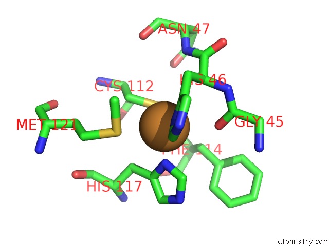
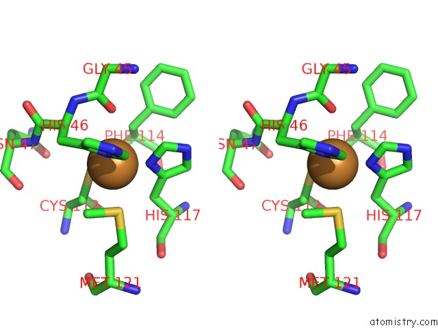
Copper binding site 1 out of 4 in 3azu
Go back to
Copper binding site 1 out
of 4 in the X-Ray Crystal Structure of the Two Site-Specific Mutants HIS35GLN and HIS35LEU of Azurin From Pseudomonas Aeruginosa
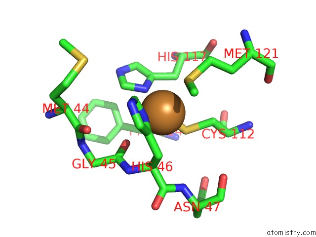
Mono view
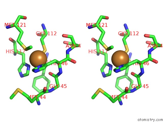
Stereo pair view
Mono view
Stereo pair view
A full contact list of Copper with other atoms in the Cu binding
site number 1 of X-Ray Crystal Structure of the Two Site-Specific Mutants HIS35GLN and HIS35LEU of Azurin From Pseudomonas Aeruginosa within 5.0Å range:
|
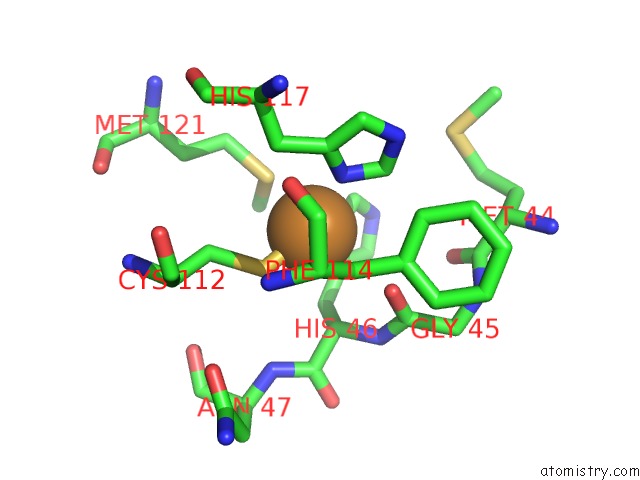
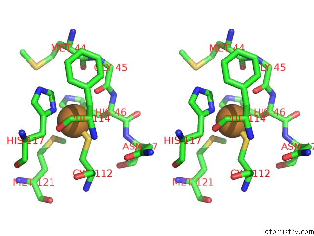
Copper binding site 2 out of 4 in 3azu
Go back to
Copper binding site 2 out
of 4 in the X-Ray Crystal Structure of the Two Site-Specific Mutants HIS35GLN and HIS35LEU of Azurin From Pseudomonas Aeruginosa
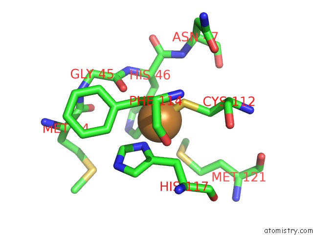
Mono view
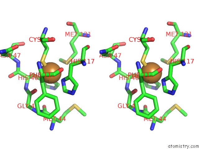
Stereo pair view
Mono view
Stereo pair view
A full contact list of Copper with other atoms in the Cu binding
site number 2 of X-Ray Crystal Structure of the Two Site-Specific Mutants HIS35GLN and HIS35LEU of Azurin From Pseudomonas Aeruginosa within 5.0Å range:
|
Copper binding site 3 out of 4 in 3azu
Go back to
Copper binding site 3 out
of 4 in the X-Ray Crystal Structure of the Two Site-Specific Mutants HIS35GLN and HIS35LEU of Azurin From Pseudomonas Aeruginosa
Mono view
Stereo pair view
Mono view
Stereo pair view
A full contact list of Copper with other atoms in the Cu binding
site number 3 of X-Ray Crystal Structure of the Two Site-Specific Mutants HIS35GLN and HIS35LEU of Azurin From Pseudomonas Aeruginosa within 5.0Å range:
|
Copper binding site 4 out of 4 in 3azu
Go back to
Copper binding site 4 out
of 4 in the X-Ray Crystal Structure of the Two Site-Specific Mutants HIS35GLN and HIS35LEU of Azurin From Pseudomonas Aeruginosa
Mono view
Stereo pair view
Mono view
Stereo pair view
A full contact list of Copper with other atoms in the Cu binding
site number 4 of X-Ray Crystal Structure of the Two Site-Specific Mutants HIS35GLN and HIS35LEU of Azurin From Pseudomonas Aeruginosa within 5.0Å range:
|
Reference:
H.Nar,
A.Messerschmidt,
R.Huber,
M.Van De Kamp,
G.W.Canters.
X-Ray Crystal Structure of the Two Site-Specific Mutants HIS35GLN and HIS35LEU of Azurin From Pseudomonas Aeruginosa. J.Mol.Biol. V. 218 427 1991.
ISSN: ISSN 0022-2836
PubMed: 1901363
DOI: 10.1016/0022-2836(91)90723-J
Page generated: Mon Jul 14 01:57:54 2025
ISSN: ISSN 0022-2836
PubMed: 1901363
DOI: 10.1016/0022-2836(91)90723-J
Last articles
Fe in 2YXOFe in 2YRS
Fe in 2YXC
Fe in 2YNM
Fe in 2YVJ
Fe in 2YP1
Fe in 2YU2
Fe in 2YU1
Fe in 2YQB
Fe in 2YOO