Copper »
PDB 2z7y-3aws »
2zmz »
Copper in PDB 2zmz: The 1.37-A Crystal Structure of the Hydroxylamine-Induced Deoxy-Form of the Copper-Bound Tyrosinase in Complex with A Caddie Protein From Streptomyces Castaneoglobisporus
Enzymatic activity of The 1.37-A Crystal Structure of the Hydroxylamine-Induced Deoxy-Form of the Copper-Bound Tyrosinase in Complex with A Caddie Protein From Streptomyces Castaneoglobisporus
All present enzymatic activity of The 1.37-A Crystal Structure of the Hydroxylamine-Induced Deoxy-Form of the Copper-Bound Tyrosinase in Complex with A Caddie Protein From Streptomyces Castaneoglobisporus:
1.14.18.1;
1.14.18.1;
Protein crystallography data
The structure of The 1.37-A Crystal Structure of the Hydroxylamine-Induced Deoxy-Form of the Copper-Bound Tyrosinase in Complex with A Caddie Protein From Streptomyces Castaneoglobisporus, PDB code: 2zmz
was solved by
Y.Matoba,
M.Sugiyama,
with X-Ray Crystallography technique. A brief refinement statistics is given in the table below:
| Resolution Low / High (Å) | 30.00 / 1.37 |
| Space group | P 21 21 2 |
| Cell size a, b, c (Å), α, β, γ (°) | 65.180, 98.030, 55.200, 90.00, 90.00, 90.00 |
| R / Rfree (%) | 18.6 / 23.2 |
Copper Binding Sites:
The binding sites of Copper atom in the The 1.37-A Crystal Structure of the Hydroxylamine-Induced Deoxy-Form of the Copper-Bound Tyrosinase in Complex with A Caddie Protein From Streptomyces Castaneoglobisporus
(pdb code 2zmz). This binding sites where shown within
5.0 Angstroms radius around Copper atom.
In total 4 binding sites of Copper where determined in the The 1.37-A Crystal Structure of the Hydroxylamine-Induced Deoxy-Form of the Copper-Bound Tyrosinase in Complex with A Caddie Protein From Streptomyces Castaneoglobisporus, PDB code: 2zmz:
Jump to Copper binding site number: 1; 2; 3; 4;
In total 4 binding sites of Copper where determined in the The 1.37-A Crystal Structure of the Hydroxylamine-Induced Deoxy-Form of the Copper-Bound Tyrosinase in Complex with A Caddie Protein From Streptomyces Castaneoglobisporus, PDB code: 2zmz:
Jump to Copper binding site number: 1; 2; 3; 4;
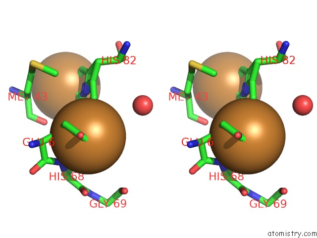
Copper binding site 1 out of 4 in 2zmz
Go back to
Copper binding site 1 out
of 4 in the The 1.37-A Crystal Structure of the Hydroxylamine-Induced Deoxy-Form of the Copper-Bound Tyrosinase in Complex with A Caddie Protein From Streptomyces Castaneoglobisporus

Mono view
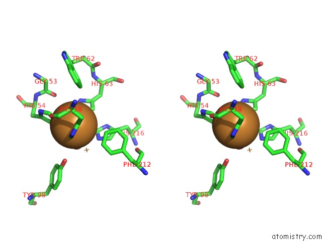
Stereo pair view
Mono view
Stereo pair view
A full contact list of Copper with other atoms in the Cu binding
site number 1 of The 1.37-A Crystal Structure of the Hydroxylamine-Induced Deoxy-Form of the Copper-Bound Tyrosinase in Complex with A Caddie Protein From Streptomyces Castaneoglobisporus within 5.0Å range:
|
Copper binding site 2 out of 4 in 2zmz
Go back to
Copper binding site 2 out
of 4 in the The 1.37-A Crystal Structure of the Hydroxylamine-Induced Deoxy-Form of the Copper-Bound Tyrosinase in Complex with A Caddie Protein From Streptomyces Castaneoglobisporus

Mono view
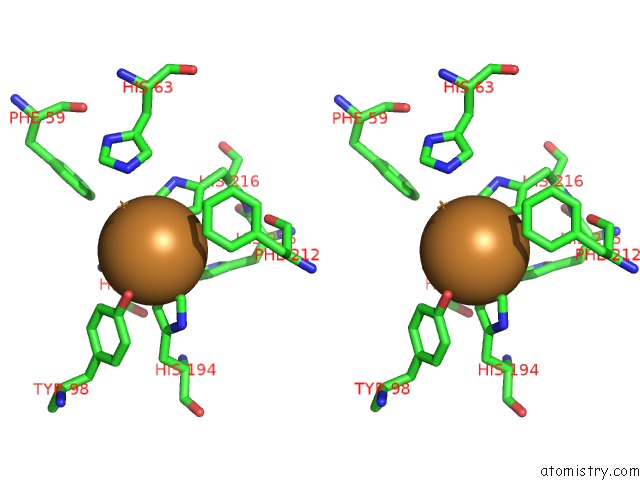
Stereo pair view
Mono view
Stereo pair view
A full contact list of Copper with other atoms in the Cu binding
site number 2 of The 1.37-A Crystal Structure of the Hydroxylamine-Induced Deoxy-Form of the Copper-Bound Tyrosinase in Complex with A Caddie Protein From Streptomyces Castaneoglobisporus within 5.0Å range:
|
Copper binding site 3 out of 4 in 2zmz
Go back to
Copper binding site 3 out
of 4 in the The 1.37-A Crystal Structure of the Hydroxylamine-Induced Deoxy-Form of the Copper-Bound Tyrosinase in Complex with A Caddie Protein From Streptomyces Castaneoglobisporus
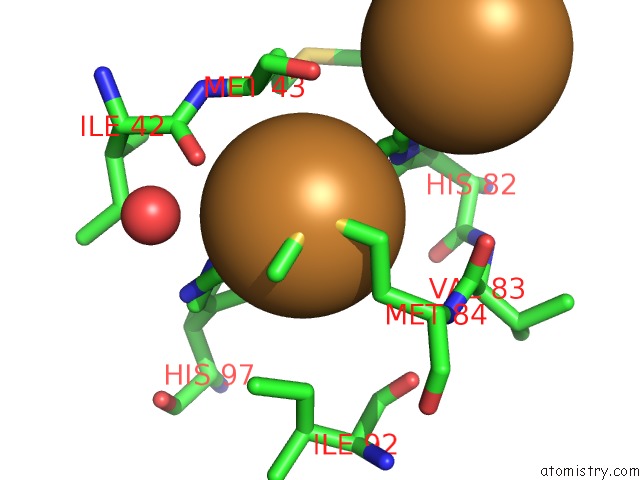
Mono view
Stereo pair view
Mono view
Stereo pair view
A full contact list of Copper with other atoms in the Cu binding
site number 3 of The 1.37-A Crystal Structure of the Hydroxylamine-Induced Deoxy-Form of the Copper-Bound Tyrosinase in Complex with A Caddie Protein From Streptomyces Castaneoglobisporus within 5.0Å range:
|
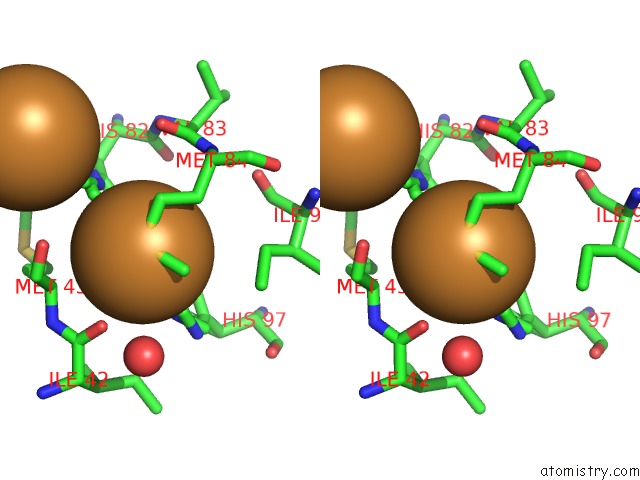
Copper binding site 4 out of 4 in 2zmz
Go back to
Copper binding site 4 out
of 4 in the The 1.37-A Crystal Structure of the Hydroxylamine-Induced Deoxy-Form of the Copper-Bound Tyrosinase in Complex with A Caddie Protein From Streptomyces Castaneoglobisporus

Mono view
Stereo pair view
Mono view
Stereo pair view
A full contact list of Copper with other atoms in the Cu binding
site number 4 of The 1.37-A Crystal Structure of the Hydroxylamine-Induced Deoxy-Form of the Copper-Bound Tyrosinase in Complex with A Caddie Protein From Streptomyces Castaneoglobisporus within 5.0Å range:
|
Reference:
Y.Matoba,
H.Yoshitsu,
H.-J.Jeon,
K.Oda,
M.Noda,
T.Kumagai,
M.Sugiyama.
X-Ray Snapshots of A Hydroxylation Mechanism of Tyrosinase To Be Published.
Page generated: Wed Jul 31 00:29:39 2024
Last articles
Zn in 9J0NZn in 9J0O
Zn in 9J0P
Zn in 9FJX
Zn in 9EKB
Zn in 9C0F
Zn in 9CAH
Zn in 9CH0
Zn in 9CH3
Zn in 9CH1