Copper »
PDB 2vr7-2xv0 »
2w88 »
Copper in PDB 2w88: Plastocyanin Variant with N-Terminal Methionine - Open Structure
Protein crystallography data
The structure of Plastocyanin Variant with N-Terminal Methionine - Open Structure, PDB code: 2w88
was solved by
P.B.Crowley,
P.M.Matias,
A.R.Khan,
with X-Ray Crystallography technique. A brief refinement statistics is given in the table below:
| Resolution Low / High (Å) | 110.43 / 2.89 |
| Space group | P 31 1 2 |
| Cell size a, b, c (Å), α, β, γ (°) | 127.700, 127.700, 94.990, 90.00, 90.00, 120.00 |
| R / Rfree (%) | 19.433 / 22.285 |
Other elements in 2w88:
The structure of Plastocyanin Variant with N-Terminal Methionine - Open Structure also contains other interesting chemical elements:
| Zinc | (Zn) | 7 atoms |
Copper Binding Sites:
The binding sites of Copper atom in the Plastocyanin Variant with N-Terminal Methionine - Open Structure
(pdb code 2w88). This binding sites where shown within
5.0 Angstroms radius around Copper atom.
In total 3 binding sites of Copper where determined in the Plastocyanin Variant with N-Terminal Methionine - Open Structure, PDB code: 2w88:
Jump to Copper binding site number: 1; 2; 3;
In total 3 binding sites of Copper where determined in the Plastocyanin Variant with N-Terminal Methionine - Open Structure, PDB code: 2w88:
Jump to Copper binding site number: 1; 2; 3;
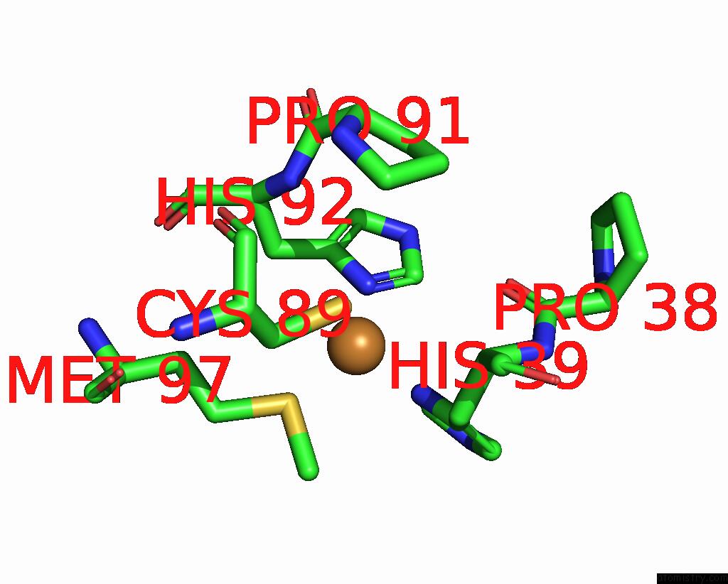
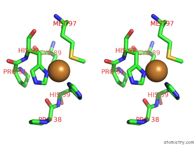
Copper binding site 1 out of 3 in 2w88
Go back to
Copper binding site 1 out
of 3 in the Plastocyanin Variant with N-Terminal Methionine - Open Structure
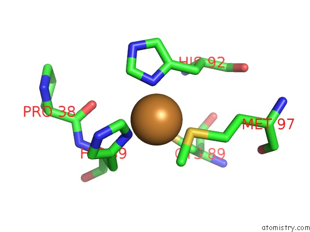
Mono view
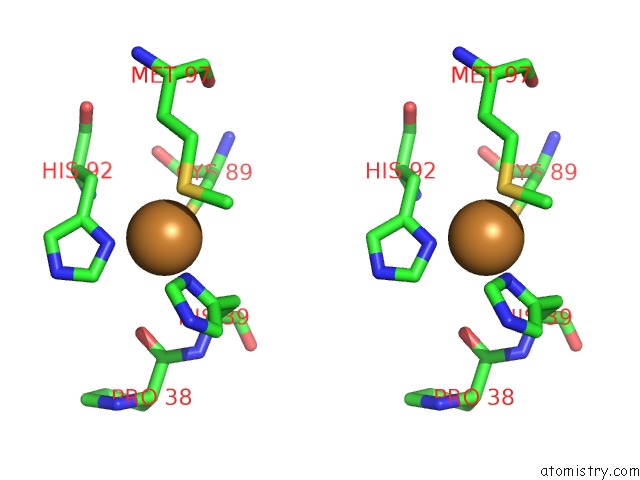
Stereo pair view
Mono view
Stereo pair view
A full contact list of Copper with other atoms in the Cu binding
site number 1 of Plastocyanin Variant with N-Terminal Methionine - Open Structure within 5.0Å range:
|
Copper binding site 2 out of 3 in 2w88
Go back to
Copper binding site 2 out
of 3 in the Plastocyanin Variant with N-Terminal Methionine - Open Structure
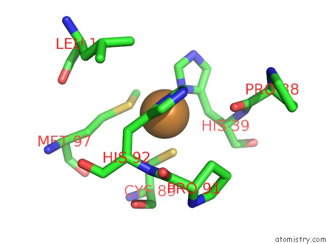
Mono view
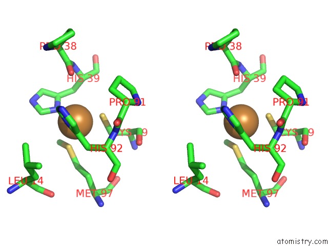
Stereo pair view
Mono view
Stereo pair view
A full contact list of Copper with other atoms in the Cu binding
site number 2 of Plastocyanin Variant with N-Terminal Methionine - Open Structure within 5.0Å range:
|
Copper binding site 3 out of 3 in 2w88
Go back to
Copper binding site 3 out
of 3 in the Plastocyanin Variant with N-Terminal Methionine - Open Structure
Mono view
Stereo pair view
Mono view
Stereo pair view
A full contact list of Copper with other atoms in the Cu binding
site number 3 of Plastocyanin Variant with N-Terminal Methionine - Open Structure within 5.0Å range:
|
Reference:
P.B.Crowley,
P.M.Matias,
A.R.Khan,
M.Roessle,
D.I.Svergun.
Metal-Mediated Self-Assembly of A Beta-Sandwich Protein. Chemistry V. 15 12672 2009.
ISSN: ISSN 0947-6539
PubMed: 19834935
DOI: 10.1002/CHEM.200901410
Page generated: Mon Jul 14 01:28:03 2025
ISSN: ISSN 0947-6539
PubMed: 19834935
DOI: 10.1002/CHEM.200901410
Last articles
F in 4IWFF in 4IXE
F in 4IW6
F in 4IVW
F in 4IVY
F in 4IVO
F in 4IV4
F in 4IVM
F in 4IV2
F in 4IUI