Copper »
PDB 2idq-2pp7 »
2plt »
Copper in PDB 2plt: Structure Determination of Plastocyanin From A Crystal Specimen with Hemihedral Twinning Fraction of One-Half
Protein crystallography data
The structure of Structure Determination of Plastocyanin From A Crystal Specimen with Hemihedral Twinning Fraction of One-Half, PDB code: 2plt
was solved by
M.R.Redinbo,
S.Merchant,
T.O.Yeates,
with X-Ray Crystallography technique. A brief refinement statistics is given in the table below:
| Resolution Low / High (Å) | N/A / 1.50 |
| Space group | P 32 |
| Cell size a, b, c (Å), α, β, γ (°) | 61.800, 61.800, 25.200, 90.00, 90.00, 120.00 |
| R / Rfree (%) | 16.8 / n/a |
Other elements in 2plt:
The structure of Structure Determination of Plastocyanin From A Crystal Specimen with Hemihedral Twinning Fraction of One-Half also contains other interesting chemical elements:
| Calcium | (Ca) | 1 atom |
Copper Binding Sites:
The binding sites of Copper atom in the Structure Determination of Plastocyanin From A Crystal Specimen with Hemihedral Twinning Fraction of One-Half
(pdb code 2plt). This binding sites where shown within
5.0 Angstroms radius around Copper atom.
In total only one binding site of Copper was determined in the Structure Determination of Plastocyanin From A Crystal Specimen with Hemihedral Twinning Fraction of One-Half, PDB code: 2plt:
In total only one binding site of Copper was determined in the Structure Determination of Plastocyanin From A Crystal Specimen with Hemihedral Twinning Fraction of One-Half, PDB code: 2plt:
Copper binding site 1 out of 1 in 2plt
Go back to
Copper binding site 1 out
of 1 in the Structure Determination of Plastocyanin From A Crystal Specimen with Hemihedral Twinning Fraction of One-Half
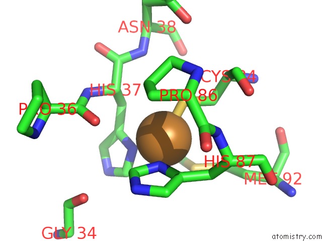
Mono view
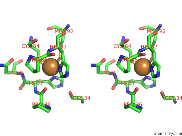
Stereo pair view
Mono view
Stereo pair view
A full contact list of Copper with other atoms in the Cu binding
site number 1 of Structure Determination of Plastocyanin From A Crystal Specimen with Hemihedral Twinning Fraction of One-Half within 5.0Å range:
|
Reference:
M.R.Redinbo,
D.Cascio,
M.K.Choukair,
D.Rice,
S.Merchant,
T.O.Yeates.
The 1.5-A Crystal Structure of Plastocyanin From the Green Alga Chlamydomonas Reinhardtii. Biochemistry V. 32 10560 1993.
ISSN: ISSN 0006-2960
PubMed: 8399201
DOI: 10.1021/BI00091A005
Page generated: Mon Jul 14 01:17:52 2025
ISSN: ISSN 0006-2960
PubMed: 8399201
DOI: 10.1021/BI00091A005
Last articles
Fe in 2YXOFe in 2YRS
Fe in 2YXC
Fe in 2YNM
Fe in 2YVJ
Fe in 2YP1
Fe in 2YU2
Fe in 2YU1
Fe in 2YQB
Fe in 2YOO